• 文献检索
  • 文档翻译
  • 深度研究
  • 学术资讯
  • Suppr Zotero 插件Zotero 插件
  • 邀请有礼
  • 套餐&价格
  • 历史记录
应用&插件
Suppr Zotero 插件Zotero 插件浏览器插件Mac 客户端Windows 客户端微信小程序
定价
高级版会员购买积分包购买API积分包
服务
文献检索文档翻译深度研究API 文档MCP 服务
关于我们
关于 Suppr公司介绍联系我们用户协议隐私条款
关注我们

Suppr 超能文献

核心技术专利:CN118964589B侵权必究
粤ICP备2023148730 号-1Suppr @ 2026

文献检索

告别复杂PubMed语法,用中文像聊天一样搜索,搜遍4000万医学文献。AI智能推荐,让科研检索更轻松。

立即免费搜索

文件翻译

保留排版,准确专业,支持PDF/Word/PPT等文件格式,支持 12+语言互译。

免费翻译文档

深度研究

AI帮你快速写综述,25分钟生成高质量综述,智能提取关键信息,辅助科研写作。

立即免费体验

两种用于测量胫股关节间隙宽度的影像学技术的比较。

Comparison of 2 Radiographic Techniques for Measurement of Tibiofemoral Joint Space Width.

作者信息

Mehta Nabil, Duryea Jeffrey, Badger Gary J, Akelman Matthew R, Jones Morgan H, Spindler Kurt P, Fleming Braden C

机构信息

Department of Orthopaedics, The Warren Alpert Medical School of Brown University/Rhode Island Hospital, Providence, Rhode Island, USA.

Department of Radiology, Brigham and Women's Hospital/Harvard University, Boston Massachusetts, USA.

出版信息

Orthop J Sports Med. 2017 Sep 26;5(9):2325967117728675. doi: 10.1177/2325967117728675. eCollection 2017 Sep.

DOI:10.1177/2325967117728675
PMID:28989937
原文链接:https://pmc.ncbi.nlm.nih.gov/articles/PMC5624356/
Abstract

BACKGROUND

No consensus is available regarding the best method for measuring tibiofemoral joint space width (JSW) on radiographs to quantify joint changes after injury. Studies that track articular cartilage thickness after injury frequently use patients' uninjured contralateral knees as controls, although the literature supporting this comparison is limited.

PURPOSE

(1) To compare JSW measurements using 2 established measurement techniques in healthy control participants and (2) to determine whether the mean JSW of the uninjured contralateral knee in a cohort with anterior cruciate ligament (ACL) reconstruction is different from that obtained from a true control population.

STUDY DESIGN

Cohort study (diagnosis); Level of evidence, 2.

METHODS

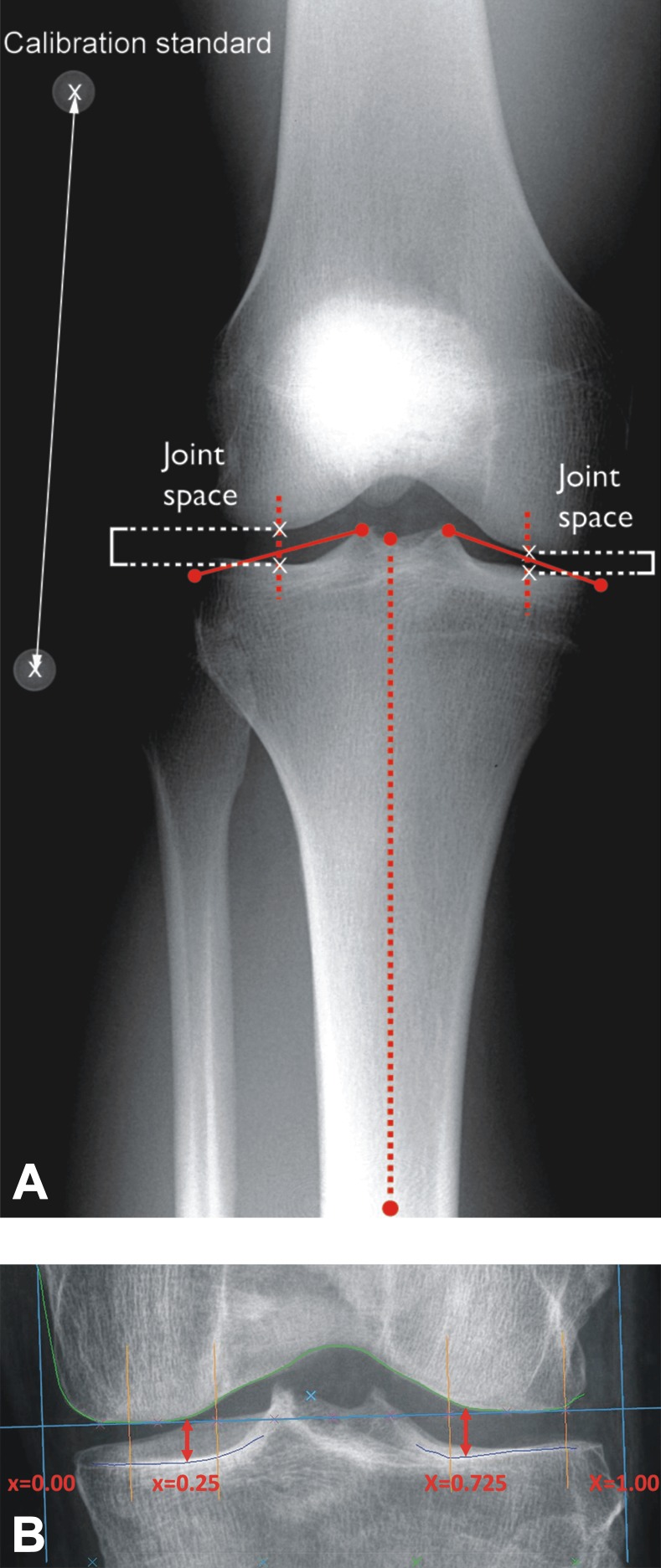
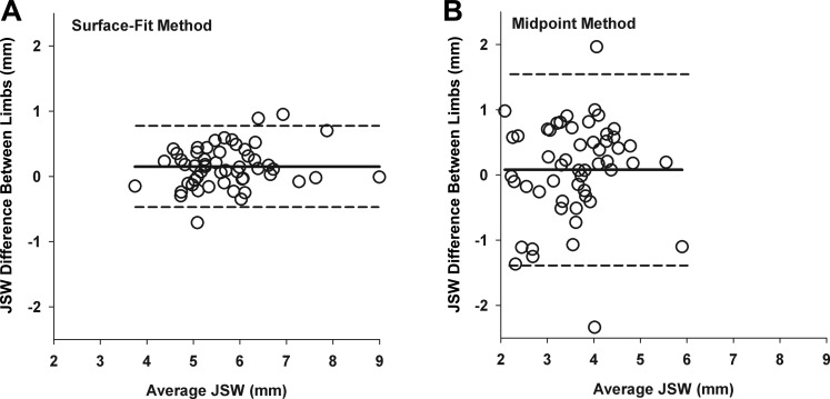
Medial and lateral JSWs were measured on standardized, bilateral, semiflexed metatarsophalangeal positioning, posteroanterior radiographs of 60 healthy individuals (26 females; mean ± SD age, 25 ± 6.2 years; no history of knee injury) via 2 published techniques: a computerized surface-delineation method (surface-fit method) and a manual digitization method (midpoint method). Bland-Altman method was used to examine the agreement between JSW measurements obtained with the 2 methods and to examine the agreement between measurements obtained on left and right knees within a participant for each measurement method. Within- and between-participant variance components and intraclass correlation coefficients (ICCs) were computed for JSW measurements corresponding to each method. Two-sample tests were used to compare the surface-fit method measurements of mean JSW of the true control group (n = 60) with the previously published mean JSW measurements from the Multicenter Orthopaedics Outcomes Network (MOON) nested cohort of 262 contralateral uninjured knees 2 to 3 years after ACL reconstruction.

RESULTS

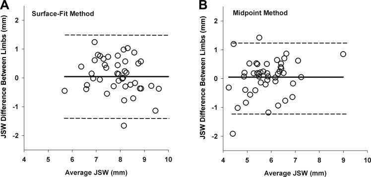
For JSW in the medial compartment, the surface-fit method had lower within-participant interknee variability (σ, 0.064; 95% CI, 0.04-0.09) compared with the midpoint method (σ, 0.28; 95% CI, 0.20-0.43) and a higher ICC (0.93 vs 0.65; < .001). Lateral JSW values were similar for the surface-fit method (σ, 0.27; 95% CI, 0.18-0.43) and the midpoint method (σ, 0.20; 95% CI, 0.14-0.31), with ICCs of 0.75 and 0.77, respectively ( = .80). With the surface-fit method, mean JSW measurements of the medial and lateral compartments of a control population were not significantly different from the contralateral uninjured knees of patients after ACL reconstruction.

CONCLUSION

For measuring medial JSW, the surface-fit method was less variable across knees within a participant than the midpoint method, as evidenced by larger ICCs and lower interknee variability. For measuring lateral JSW, the 2 methods were similar. The JSW measurements of uninjured contralateral knees of patients with ACL reconstruction at 2 to 3 years postsurgery were not significantly different from those of a cohort of healthy control participants. Future work should be performed to demonstrate the validity of these methods for documenting change over time in the ACL-reconstructed knee.

摘要

背景

关于在X线片上测量胫股关节间隙宽度(JSW)以量化损伤后关节变化的最佳方法,目前尚无共识。追踪损伤后关节软骨厚度的研究经常将患者未受伤的对侧膝关节作为对照,尽管支持这种比较的文献有限。

目的

(1)在健康对照参与者中比较使用两种既定测量技术测量JSW的情况,(2)确定前交叉韧带(ACL)重建队列中未受伤对侧膝关节的平均JSW是否与真正对照组人群的平均JSW不同。

研究设计

队列研究(诊断);证据等级,2级。

方法

通过两种已发表的技术,在60名健康个体(26名女性;平均±标准差年龄,25±6.2岁;无膝关节损伤史)的标准化、双侧、半屈曲跖趾关节定位的正位X线片上测量内侧和外侧JSW:一种计算机表面描绘方法(表面拟合方法)和一种手动数字化方法(中点法)。采用Bland-Altman方法检查两种方法获得的JSW测量值之间的一致性,以及在每个测量方法中参与者左右膝关节测量值之间的一致性。计算每种方法对应的JSW测量值的参与者内和参与者间方差分量以及组内相关系数(ICC)。采用两样本检验比较真正对照组(n = 60)的表面拟合方法测量的平均JSW与多中心骨科结果网络(MOON)嵌套队列中262例ACL重建术后2至3年对侧未受伤膝关节先前发表的平均JSW测量值。

结果

在内侧间室的JSW测量中,与中点法(σ,0.28;95%CI,0.20 - 0.43)相比,表面拟合方法的参与者内膝关节间变异性较低(σ,0.064;95%CI,0.04 - 0.09),ICC较高(0.93对0.65;P <.001)。外侧JSW值在表面拟合方法(σ,0.27;95%CI,0.18 - 0.43)和中点法(σ,0.20;95%CI,0.14 - 0.31)中相似,ICC分别为0.75和0.77(P =.80)。采用表面拟合方法时,对照组人群内侧和外侧间室的平均JSW测量值与ACL重建术后患者的对侧未受伤膝关节无显著差异。

结论

对于测量内侧JSW,表面拟合方法在参与者内膝关节间的变异性低于中点法,这由更高的ICC和更低的膝关节间变异性证明。对于测量外侧JSW,两种方法相似。ACL重建术后2至3年患者未受伤对侧膝关节的JSW测量值与健康对照参与者队列的测量值无显著差异。未来应开展工作以证明这些方法记录ACL重建膝关节随时间变化的有效性。

https://cdn.ncbi.nlm.nih.gov/pmc/blobs/191a/5624356/207ce3949c88/10.1177_2325967117728675-fig5.jpg
https://cdn.ncbi.nlm.nih.gov/pmc/blobs/191a/5624356/a4fa10e267d8/10.1177_2325967117728675-fig1.jpg
https://cdn.ncbi.nlm.nih.gov/pmc/blobs/191a/5624356/58682a7d6f3b/10.1177_2325967117728675-fig2.jpg
https://cdn.ncbi.nlm.nih.gov/pmc/blobs/191a/5624356/e31527e93490/10.1177_2325967117728675-fig3.jpg
https://cdn.ncbi.nlm.nih.gov/pmc/blobs/191a/5624356/9699d6bf4a97/10.1177_2325967117728675-fig4.jpg
https://cdn.ncbi.nlm.nih.gov/pmc/blobs/191a/5624356/207ce3949c88/10.1177_2325967117728675-fig5.jpg

相似文献

1
Comparison of 2 Radiographic Techniques for Measurement of Tibiofemoral Joint Space Width.两种用于测量胫股关节间隙宽度的影像学技术的比较。
Orthop J Sports Med. 2017 Sep 26;5(9):2325967117728675. doi: 10.1177/2325967117728675. eCollection 2017 Sep.
2
Assessment of early tibiofemoral joint space width changes after anterior cruciate ligament injury and reconstruction: a matched case-control study.前交叉韧带损伤及重建后早期胫股关节间隙宽度变化的评估:一项匹配病例对照研究。
Am J Sports Med. 2013 Apr;41(4):769-78. doi: 10.1177/0363546513477838. Epub 2013 Mar 4.
3
Early radiographic features in patients with anterior cruciate ligament rupture.前交叉韧带断裂患者的早期影像学特征。
Ann Rheum Dis. 2000 Aug;59(8):641-6. doi: 10.1136/ard.59.8.641.
4
Measurement of radiographic joint space width in the tibiofemoral compartment of the osteoarthritic knee: comparison of standing anteroposterior and Lyon schuss views.骨关节炎膝关节胫股关节间隙的X线关节间隙宽度测量:站立前后位与里昂切线位的比较
Arthritis Rheum. 2003 Feb;48(2):378-84. doi: 10.1002/art.10773.
5
Differences in the Lateral Compartment Joint Space Width After Anterior Cruciate Ligament Reconstruction: Data From the MOON Onsite Cohort.前交叉韧带重建后外侧间室关节间隙宽度的差异:来自 MOON 现场队列的数据。
Am J Sports Med. 2018 Mar;46(4):876-882. doi: 10.1177/0363546517751139. Epub 2018 Feb 2.
6
Reproducibility of the semiflexed (metatarsophalangeal) radiographic knee position and automated measurements of medial tibiofemoral joint space width in a multicenter clinical trial of knee osteoarthritis.在一项膝关节骨关节炎多中心临床试验中,半屈曲(跖趾关节)X线膝关节位的可重复性及胫股内侧关节间隙宽度的自动测量
J Rheumatol. 2004 Aug;31(8):1588-97.
7
Relationship between markers of type II collagen metabolism and tibiofemoral joint space width changes after ACL injury and reconstruction.ACL 损伤与重建后Ⅱ型胶原代谢标志物与胫股关节间隙宽度变化的关系。
Am J Sports Med. 2013 Apr;41(4):779-87. doi: 10.1177/0363546513476481. Epub 2013 Feb 19.
8
Pitfalls in the accurate measurement of joint space narrowing in semiflexed, anteroposterior radiographic imaging of the knee.膝关节半屈曲前后位X线成像中关节间隙狭窄精确测量的陷阱。
Arthritis Rheum. 2004 Aug;50(8):2508-15. doi: 10.1002/art.20363.
9
Meniscus treatment and age associated with narrower radiographic joint space width 2-3 years after ACL reconstruction: data from the MOON onsite cohort.前交叉韧带重建术后2 - 3年,半月板治疗及年龄与影像学显示的关节间隙宽度变窄相关:来自MOON现场队列的数据
Osteoarthritis Cartilage. 2015 Apr;23(4):581-8. doi: 10.1016/j.joca.2014.12.018. Epub 2015 Jan 3.
10
Relationships Between Tibiofemoral Contact Forces and Cartilage Morphology at 2 to 3 Years After Single-Bundle Hamstring Anterior Cruciate Ligament Reconstruction and in Healthy Knees.单束腘绳肌前交叉韧带重建术后2至3年时与健康膝关节中胫股接触力和软骨形态之间的关系。
Orthop J Sports Med. 2017 Aug 31;5(8):2325967117722506. doi: 10.1177/2325967117722506. eCollection 2017 Aug.

引用本文的文献

1
Plain Radiographs Have Limited Utility in the Evaluation of Discoid Lateral Meniscus.普通X线片在盘状外侧半月板评估中的应用有限。
J Pediatr Soc North Am. 2024 Feb 5;5(4):756. doi: 10.55275/JPOSNA-2023-756. eCollection 2023 Nov.
2
Stress radiography of medial knee instability provides a reliable correlation with the severity of injury and medial joint space opening-A robotic biomechanical cadaveric study.膝关节内侧不稳定的应力放射成像与损伤严重程度及内侧关节间隙增宽具有可靠的相关性——一项机器人生物力学尸体研究。
Knee Surg Sports Traumatol Arthrosc. 2025 Jul;33(7):2438-2446. doi: 10.1002/ksa.12594. Epub 2025 Jan 22.
3

本文引用的文献

1
Effect of Matching or Overconstraining Knee Laxity During Anterior Cruciate Ligament Reconstruction on Knee Osteoarthritis and Clinical Outcomes: A Randomized Controlled Trial With 84-Month Follow-up.前交叉韧带重建术中匹配或过度约束膝关节松弛度对膝骨关节炎及临床结局的影响:一项长达84个月随访的随机对照试验
Am J Sports Med. 2016 Jul;44(7):1660-70. doi: 10.1177/0363546516638387. Epub 2016 Apr 19.
2
Comparison of radiographic joint space width and magnetic resonance imaging for prediction of knee replacement: A longitudinal case-control study from the Osteoarthritis Initiative.X线片关节间隙宽度与磁共振成像预测膝关节置换的比较:来自骨关节炎倡议组织的一项纵向病例对照研究。
Eur Radiol. 2016 Jun;26(6):1942-51. doi: 10.1007/s00330-015-3977-8. Epub 2015 Sep 16.
3
Clinical and Radiological Outcomes After Lateral Meniscal Allograft Transplantation Through a Medial Arthrotomy: A Minimum 4-Year Follow-up.
经内侧关节切开术行外侧半月板同种异体移植后的临床及影像学结果:至少4年随访
Orthop J Sports Med. 2025 Jan 16;13(1):23259671241306431. doi: 10.1177/23259671241306431. eCollection 2025 Jan.
4
Joint line and knee osteotomy.关节线与膝关节截骨术
EFORT Open Rev. 2024 May 10;9(5):375-386. doi: 10.1530/EOR-24-0037.
5
Valgus stress radiographs have a minor additional value in detecting lateral cartilage wear when selecting patients for medial partial knee replacement.在选择内侧部分膝关节置换术患者时,外翻应力量线片在检测外侧软骨磨损方面具有较小的附加价值。
Arch Orthop Trauma Surg. 2024 Apr;144(4):1721-1732. doi: 10.1007/s00402-023-05187-0. Epub 2024 Jan 12.
6
Early knee OA definition-what do we know at this stage? An imaging perspective.早期膝关节骨关节炎的定义——现阶段我们了解多少?影像学视角。
Ther Adv Musculoskelet Dis. 2023 Mar 14;15:1759720X231158204. doi: 10.1177/1759720X231158204. eCollection 2023.
7
Medial joint space narrowing progresses after pullout repair of medial meniscus posterior root tear.内侧半月板后根撕裂的拉出修复术后,内侧关节间隙变窄会进展。
Int Orthop. 2023 Oct;47(10):2401-2407. doi: 10.1007/s00264-023-05701-4. Epub 2023 Jan 30.
8
Planning Results for High Tibial Osteotomies in Degenerative Varus Osteoarthritis Using Standing and Supine Whole Leg Radiographs.使用站立位和仰卧位全下肢正位片规划胫骨高位截骨术治疗退行性内翻型骨关节炎的结果。
Orthop Surg. 2021 Feb;13(1):77-82. doi: 10.1111/os.12833. Epub 2020 Nov 30.
9
A quantitative metric for knee osteoarthritis: reference values of joint space loss.膝关节骨关节炎的定量指标:关节间隙损失的参考值。
Osteoarthritis Cartilage. 2018 Sep;26(9):1215-1224. doi: 10.1016/j.joca.2018.05.014. Epub 2018 May 26.
Risk Factors and Predictors of Subsequent ACL Injury in Either Knee After ACL Reconstruction: Prospective Analysis of 2488 Primary ACL Reconstructions From the MOON Cohort.前交叉韧带重建术后任一膝关节发生后续前交叉韧带损伤的危险因素及预测因素:来自MOON队列的2488例初次前交叉韧带重建的前瞻性分析
Am J Sports Med. 2015 Jul;43(7):1583-90. doi: 10.1177/0363546515578836. Epub 2015 Apr 21.
4
Brief report: symmetricity of radiographic and MRI-detected structural joint damage in persons with knee pain--the Joints on Glucosamine (JOG) Study.简短报告:膝关节疼痛患者中X线和MRI检测到的关节结构损伤的对称性——氨基葡萄糖关节研究(JOG研究)
Osteoarthritis Cartilage. 2015 Aug;23(8):1343-7. doi: 10.1016/j.joca.2015.02.169. Epub 2015 Mar 6.
5
Meniscus treatment and age associated with narrower radiographic joint space width 2-3 years after ACL reconstruction: data from the MOON onsite cohort.前交叉韧带重建术后2 - 3年,半月板治疗及年龄与影像学显示的关节间隙宽度变窄相关:来自MOON现场队列的数据
Osteoarthritis Cartilage. 2015 Apr;23(4):581-8. doi: 10.1016/j.joca.2014.12.018. Epub 2015 Jan 3.
6
Changes to the articular cartilage thickness profile of the tibia following anterior cruciate ligament injury.前交叉韧带损伤后胫骨关节软骨厚度轮廓的变化。
Osteoarthritis Cartilage. 2014 Oct;22(10):1453-60. doi: 10.1016/j.joca.2014.06.025.
7
Milk consumption and progression of medial tibiofemoral knee osteoarthritis: data from the Osteoarthritis Initiative.牛奶摄入与膝内侧胫骨股骨骨关节炎进展的关系:来自骨关节炎倡议的数据。
Arthritis Care Res (Hoboken). 2014 Jun;66(6):802-9. doi: 10.1002/acr.22297.
8
Assessment of early tibiofemoral joint space width changes after anterior cruciate ligament injury and reconstruction: a matched case-control study.前交叉韧带损伤及重建后早期胫股关节间隙宽度变化的评估:一项匹配病例对照研究。
Am J Sports Med. 2013 Apr;41(4):769-78. doi: 10.1177/0363546513477838. Epub 2013 Mar 4.
9
The effect of initial graft tension after anterior cruciate ligament reconstruction: a randomized clinical trial with 36-month follow-up.前交叉韧带重建后初始移植物张力的影响:一项 36 个月随访的随机临床试验。
Am J Sports Med. 2013 Jan;41(1):25-34. doi: 10.1177/0363546512464200. Epub 2012 Nov 9.
10
Incidence of contralateral and ipsilateral anterior cruciate ligament (ACL) injury after primary ACL reconstruction and return to sport.初次前交叉韧带(ACL)重建术后和重返运动后对侧和同侧 ACL 损伤的发生率。
Clin J Sport Med. 2012 Mar;22(2):116-21. doi: 10.1097/JSM.0b013e318246ef9e.