Department of pathology, Xuzhou Medical University, Xuzhou, 221002, China.
Department of Interventional Radiology, Jining No.1 People's Hospital, Jining, Shandong Province, China.
J Exp Clin Cancer Res. 2017 Oct 17;36(1):146. doi: 10.1186/s13046-017-0610-5.
Abnormal proliferation is significantly associated with the promotion of malignant tumor. Growing evidence suggest that the signal pathways of p21-activated kinase 5 (PAK5) have been found in various tumor progression, however, the role of PAK5 in breast cancer remains largely unclear.
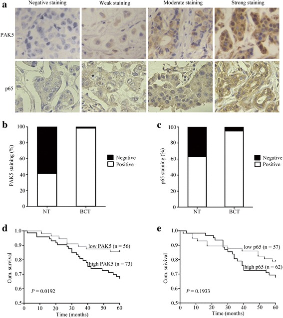
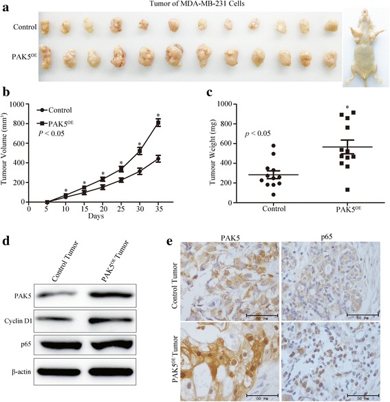
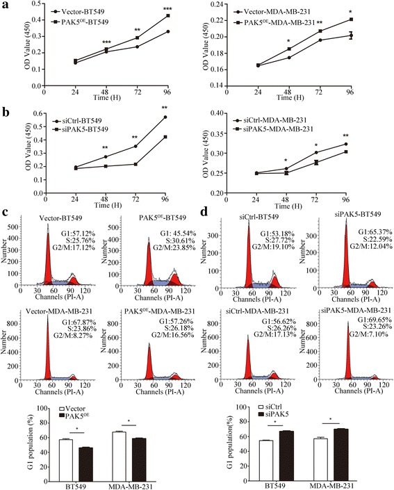
We evaluated PAK5 and p65 staining in breast cancer tissues (BCTs) and paired non-cancerous tissues (NTs) using tissue microarray (TMA) technology. The functions of PAK5 were studied in vitro and in vivo. Cell Counting Kit-8 (CCK-8) and flow cytometry were performed to determine proliferation of breast cancer cells. Phosphorylation assay and co-immunoprecipitation (co-IP) were employed to identify the regulation mechanism of p65 by PAK5. The activation of Cyclin D1 promoter was measured with luciferase reporter assay. Xenograft models in nude mice were established to explore the roles of PAK5 in breast cancer growth.
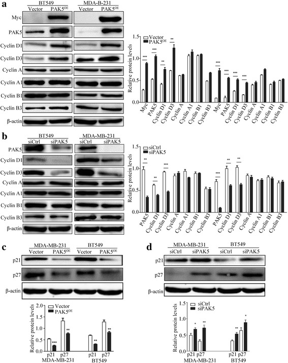
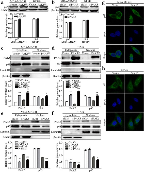
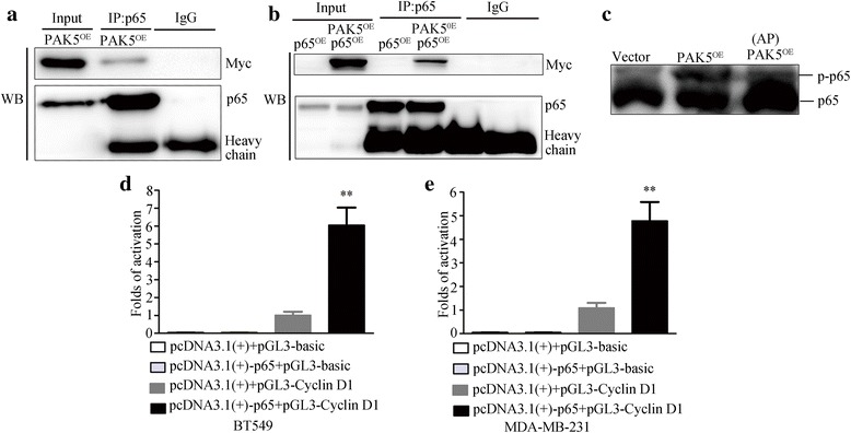
In this study, we show that PAK5 is highly expressed in breast cancer tissues and the increased PAK5 is significantly associated with breast cancer progression. Overexpression of PAK5 promotes the proliferation and cell-cycle progression by increasing the expression of Cyclin D1 in vitro and in vivo. Mechanistic studies demonstrated that PAK5 can promote the phosphorylation and the nuclear translocation of p65 subunit of nuclear factor-kappaB (NF-κB). Furthermore, p65 can directly bind to the promoter of Cyclin D1 and mediate an increase in its protein expression.
Taken together, our findings suggest that PAK5 may serve as a potential prognosis marker and therapeutic target for human breast cancer.
异常增殖与恶性肿瘤的促进显著相关。越来越多的证据表明,p21 激活激酶 5(PAK5)的信号通路已在各种肿瘤进展中被发现,然而,PAK5 在乳腺癌中的作用在很大程度上仍不清楚。
我们使用组织微阵列(TMA)技术评估了乳腺癌组织(BCT)和配对的非癌组织(NT)中的 PAK5 和 p65 染色。在体外和体内研究了 PAK5 的功能。使用细胞计数试剂盒-8(CCK-8)和流式细胞术来确定乳腺癌细胞的增殖。进行磷酸化测定和免疫共沉淀(co-IP)以鉴定 PAK5 对 p65 的调节机制。使用荧光素酶报告基因测定测量 Cyclin D1 启动子的激活。建立裸鼠异种移植模型以探索 PAK5 在乳腺癌生长中的作用。
在这项研究中,我们表明 PAK5 在乳腺癌组织中高度表达,并且增加的 PAK5 与乳腺癌的进展显著相关。PAK5 的过表达通过增加体外和体内的 Cyclin D1 表达促进增殖和细胞周期进程。机制研究表明,PAK5 可以促进核因子-κB(NF-κB)p65 亚基的磷酸化和核转位。此外,p65 可以直接结合 Cyclin D1 启动子并介导其蛋白表达增加。
总之,我们的研究结果表明,PAK5 可能作为人类乳腺癌的潜在预后标志物和治疗靶标。