• 文献检索
  • 文档翻译
  • 深度研究
  • 学术资讯
  • Suppr Zotero 插件Zotero 插件
  • 邀请有礼
  • 套餐&价格
  • 历史记录
应用&插件
Suppr Zotero 插件Zotero 插件浏览器插件Mac 客户端Windows 客户端微信小程序
定价
高级版会员购买积分包购买API积分包
服务
文献检索文档翻译深度研究API 文档MCP 服务
关于我们
关于 Suppr公司介绍联系我们用户协议隐私条款
关注我们

Suppr 超能文献

核心技术专利:CN118964589B侵权必究
粤ICP备2023148730 号-1Suppr @ 2026

文献检索

告别复杂PubMed语法,用中文像聊天一样搜索,搜遍4000万医学文献。AI智能推荐,让科研检索更轻松。

立即免费搜索

文件翻译

保留排版,准确专业,支持PDF/Word/PPT等文件格式,支持 12+语言互译。

免费翻译文档

深度研究

AI帮你快速写综述,25分钟生成高质量综述,智能提取关键信息,辅助科研写作。

立即免费体验

基于定量计算机断层扫描的有限元分析的扫描分辨率和元素大小对牛椎体力学参数的影响。

Effects of Scan Resolutions and Element Sizes on Bovine Vertebral Mechanical Parameters from Quantitative Computed Tomography-Based Finite Element Analysis.

机构信息

Department of Engineering Mechanics, Nanling Campus, Jilin University, Changchun 130025, China.

Department of Radiology, The First Hospital of Jilin University, Changchun 130021, China.

出版信息

J Healthc Eng. 2017;2017:5707568. doi: 10.1155/2017/5707568. Epub 2017 Jun 1.

DOI:10.1155/2017/5707568
PMID:29065624
原文链接:https://pmc.ncbi.nlm.nih.gov/articles/PMC5474284/
Abstract

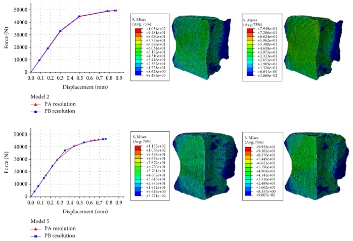
Quantitative computed tomography-based finite element analysis (QCT/FEA) has been developed to predict vertebral strength. However, QCT/FEA models may be different with scan resolutions and element sizes. The aim of this study was to explore the effects of scan resolutions and element sizes on QCT/FEA outcomes. Nine bovine vertebral bodies were scanned using the clinical CT scanner and reconstructed from datasets with the two-slice thickness, that is, 0.6 mm (PA resolution) and 1 mm (PB resolution). There were significantly linear correlations between the predicted and measured principal strains ( > 0.7, < 0.0001), and the predicted vertebral strength and stiffness were modestly correlated with the experimental values ( > 0.6, < 0.05). Two different resolutions and six different element sizes were combined in pairs, and finite element (FE) models of bovine vertebral cancellous bones in the 12 cases were obtained. It showed that the mechanical parameters of FE models with the PB resolution were similar to those with the PA resolution. The computational accuracy of FE models with the element sizes of 0.41 × 0.41 × 0.6 mm and 0.41 × 0.41 × 1 mm was higher by comparing the apparent elastic modulus and yield strength. Therefore, scan resolution and element size should be chosen optimally to improve the accuracy of QCT/FEA.

摘要

基于定量计算机断层扫描的有限元分析(QCT/FEA)已经被开发出来用于预测椎体强度。然而,QCT/FEA 模型可能会因扫描分辨率和单元大小而有所不同。本研究旨在探讨扫描分辨率和单元大小对 QCT/FEA 结果的影响。使用临床 CT 扫描仪对 9 个牛椎体进行扫描,并从数据集重建,数据集的厚度为 2 层,即 0.6mm(PA 分辨率)和 1mm(PB 分辨率)。预测的和测量的主应变之间存在显著的线性相关性( > 0.7, < 0.0001),预测的椎体强度和刚度与实验值呈中度相关( > 0.6, < 0.05)。两种不同的分辨率和六种不同的单元大小组合成对,获得了 12 例牛松质骨的有限元(FE)模型。结果表明,PB 分辨率的 FE 模型的力学参数与 PA 分辨率的 FE 模型相似。通过比较表观弹性模量和屈服强度,单元大小为 0.41×0.41×0.6mm 和 0.41×0.41×1mm 的 FE 模型的计算精度更高。因此,为了提高 QCT/FEA 的准确性,应选择最佳的扫描分辨率和单元大小。

https://cdn.ncbi.nlm.nih.gov/pmc/blobs/43e7/5474284/5f88d390db94/JHE2017-5707568.008.jpg
https://cdn.ncbi.nlm.nih.gov/pmc/blobs/43e7/5474284/a0c7c14f3edc/JHE2017-5707568.001.jpg
https://cdn.ncbi.nlm.nih.gov/pmc/blobs/43e7/5474284/bb3a3a9dcc25/JHE2017-5707568.002.jpg
https://cdn.ncbi.nlm.nih.gov/pmc/blobs/43e7/5474284/e6e2469360bc/JHE2017-5707568.003.jpg
https://cdn.ncbi.nlm.nih.gov/pmc/blobs/43e7/5474284/de93d6e94ca7/JHE2017-5707568.004.jpg
https://cdn.ncbi.nlm.nih.gov/pmc/blobs/43e7/5474284/9774ad78bd27/JHE2017-5707568.005.jpg
https://cdn.ncbi.nlm.nih.gov/pmc/blobs/43e7/5474284/826bcb06d28f/JHE2017-5707568.006.jpg
https://cdn.ncbi.nlm.nih.gov/pmc/blobs/43e7/5474284/a1bd74b094ce/JHE2017-5707568.007.jpg
https://cdn.ncbi.nlm.nih.gov/pmc/blobs/43e7/5474284/5f88d390db94/JHE2017-5707568.008.jpg

相似文献

1
Effects of Scan Resolutions and Element Sizes on Bovine Vertebral Mechanical Parameters from Quantitative Computed Tomography-Based Finite Element Analysis.基于定量计算机断层扫描的有限元分析的扫描分辨率和元素大小对牛椎体力学参数的影响。
J Healthc Eng. 2017;2017:5707568. doi: 10.1155/2017/5707568. Epub 2017 Jun 1.
2
Influence of 3D QCT scan protocol on the QCT-based finite element models of human vertebral cancellous bone.三维定量CT扫描协议对基于定量CT的人体椎体松质骨有限元模型的影响。
Med Eng Phys. 2014 Aug;36(8):1069-73. doi: 10.1016/j.medengphy.2014.05.001. Epub 2014 Jun 2.
3
The effect of in situ/in vitro three-dimensional quantitative computed tomography image voxel size on the finite element model of human vertebral cancellous bone.原位/体外三维定量计算机断层扫描图像体素大小对人椎体松质骨有限元模型的影响。
Proc Inst Mech Eng H. 2014 Nov;228(11):1208-13. doi: 10.1177/0954411914558654.
4
Quantitative Computed Tomography Protocols Affect Material Mapping and Quantitative Computed Tomography-Based Finite-Element Analysis Predicted Stiffness.定量计算机断层扫描协议影响材料映射以及基于定量计算机断层扫描的有限元分析预测的刚度。
J Biomech Eng. 2016 Sep 1;138(9):0910031-7. doi: 10.1115/1.4034172.
5
Finite element models predict in vitro vertebral body compressive strength better than quantitative computed tomography.有限元模型在预测体外椎体抗压强度方面比定量计算机断层扫描表现更好。
Bone. 2003 Oct;33(4):744-50. doi: 10.1016/s8756-3282(03)00210-2.
6
Prediction of bone strength by μCT and MDCT-based finite-element-models: how much spatial resolution is needed?基于 μCT 和 MDCT 的有限元模型预测骨强度:需要多少空间分辨率?
Eur J Radiol. 2014 Jan;83(1):e36-42. doi: 10.1016/j.ejrad.2013.10.024. Epub 2013 Nov 8.
7
Finite element modeling of the human thoracolumbar spine.人体胸腰椎的有限元建模
Spine (Phila Pa 1976). 2003 Mar 15;28(6):559-65. doi: 10.1097/01.BRS.0000049923.27694.47.
8
Finite element analyses of human vertebral bodies embedded in polymethylmethalcrylate or loaded via the hyperelastic intervertebral disc models provide equivalent predictions of experimental strength.人体椎体嵌入聚甲基丙烯酸甲酯或通过超弹性椎间盘模型加载的有限元分析提供了与实验强度相当的预测。
J Biomech. 2014 Jul 18;47(10):2512-6. doi: 10.1016/j.jbiomech.2014.04.015. Epub 2014 Apr 16.
9
Density-Dependent Material and Failure Criteria Equations Highly Affect the Accuracy and Precision of QCT/FEA-Based Predictions of Osteoporotic Vertebral Fracture Properties.密度相关的材料和失效准则方程极大地影响了基于 QCT/FEA 的骨质疏松性椎体骨折特性预测的准确性和精密度。
Ann Biomed Eng. 2021 Feb;49(2):663-672. doi: 10.1007/s10439-020-02595-w. Epub 2020 Aug 20.
10
Noninvasive prediction of vertebral body compressive strength using nonlinear finite element method and an image based technique.使用非线性有限元方法和基于图像的技术无创预测椎体压缩强度。
Phys Med. 2010 Apr;26(2):88-97. doi: 10.1016/j.ejmp.2009.08.002. Epub 2009 Sep 24.

引用本文的文献

1
Patient-Specific Bone Multiscale Modelling, Fracture Simulation and Risk Analysis-A Survey.个性化骨骼多尺度建模、骨折模拟与风险分析——综述
Materials (Basel). 2019 Dec 24;13(1):106. doi: 10.3390/ma13010106.

本文引用的文献

1
Quantitative Computed Tomography Protocols Affect Material Mapping and Quantitative Computed Tomography-Based Finite-Element Analysis Predicted Stiffness.定量计算机断层扫描协议影响材料映射以及基于定量计算机断层扫描的有限元分析预测的刚度。
J Biomech Eng. 2016 Sep 1;138(9):0910031-7. doi: 10.1115/1.4034172.
2
Automated finite element meshing of the lumbar spine: Verification and validation with 18 specimen-specific models.腰椎的自动有限元网格划分:通过18个特定标本模型进行验证与确认
J Biomech. 2016 Sep 6;49(13):2669-2676. doi: 10.1016/j.jbiomech.2016.05.025. Epub 2016 May 31.
3
Residual lifetime and 10 year absolute risks of osteoporotic fractures in Chinese men and women.
中国男性和女性骨质疏松性骨折的剩余寿命及10年绝对风险。
Curr Med Res Opin. 2015 Jun;31(6):1149-56. doi: 10.1185/03007995.2015.1037729. Epub 2015 May 20.
4
The effect of in situ/in vitro three-dimensional quantitative computed tomography image voxel size on the finite element model of human vertebral cancellous bone.原位/体外三维定量计算机断层扫描图像体素大小对人椎体松质骨有限元模型的影响。
Proc Inst Mech Eng H. 2014 Nov;228(11):1208-13. doi: 10.1177/0954411914558654.
5
Quantitative computed tomography-based finite element analysis predictions of femoral strength and stiffness depend on computed tomography settings.基于定量计算机断层扫描的股骨强度和刚度有限元分析预测取决于计算机断层扫描设置。
J Biomech. 2015 Jan 2;48(1):153-61. doi: 10.1016/j.jbiomech.2014.09.016. Epub 2014 Sep 28.
6
Specimen-specific nonlinear finite element modeling to predict vertebrae fracture loads after vertebroplasty.用于预测椎体成形术后椎体骨折负荷的特定标本非线性有限元建模。
Spine (Phila Pa 1976). 2014 Oct 15;39(22):E1291-6. doi: 10.1097/BRS.0000000000000540.
7
Influence of 3D QCT scan protocol on the QCT-based finite element models of human vertebral cancellous bone.三维定量CT扫描协议对基于定量CT的人体椎体松质骨有限元模型的影响。
Med Eng Phys. 2014 Aug;36(8):1069-73. doi: 10.1016/j.medengphy.2014.05.001. Epub 2014 Jun 2.
8
Clinical versus pre-clinical FE models for vertebral body strength predictions.临床与临床前 FE 模型在预测椎体强度方面的比较。
J Mech Behav Biomed Mater. 2014 May;33:76-83. doi: 10.1016/j.jmbbm.2012.11.018. Epub 2012 Dec 20.
9
The effect of voxel size on high-resolution peripheral computed tomography measurements of trabecular and cortical bone microstructure.体素大小对高分辨率外周计算机断层扫描测量骨小梁和皮质骨微观结构的影响。
Med Phys. 2012 Apr;39(4):1893-903. doi: 10.1118/1.3689813.
10
Prediction of the vertebral strength using a finite element model derived from low-dose biplanar imaging: benefits of subject-specific material properties.使用源于低剂量双平面成像的有限元模型预测椎体强度:基于个体的材料特性的优势。
Spine (Phila Pa 1976). 2012 Feb 1;37(3):E156-62. doi: 10.1097/BRS.0b013e3182293628.