David Juliet, Nandakumar Athira, Muniroh Muflihatul, Akiba Suminori, Yamamoto Megumi, Koriyama Chihaya
Department of Epidemiology and Preventive Medicine, Kagoshima University Graduate School of Medical and Dental Sciences, 8-35-1 Sakuragaoka, Kagoshima, 890-8544, Japan.
Department of Physiology, Faculty of Medicine, Diponegoro University, Tembalang, Semarang, 50725, Indonesia.
Eur J Med Res. 2017 Nov 9;22(1):45. doi: 10.1186/s40001-017-0287-4.
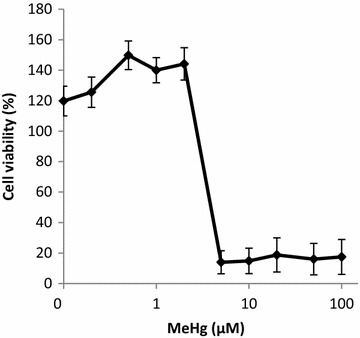
The aim of this study is to examine the inflammatory-cytokine expressions in the presence of non-cytotoxic dose of methylmercury (MeHg) in murine macrophages, which is suspected to play an important role in brain damage caused by MeHg exposure. We focused on murine macrophage inflammatory protein-2 (MIP-2), keratinocyte chemoattractant (KC), and monocyte chemoattractant protein-5 (MCP-5). MIP-2 and KC are murine functional homologues of human IL-8 and MCP-5 for human MCP-1. Furthermore, we examined the suppressive effect of N-acetyl-L-cysteine (NAC) on the MeHg-induced inflammatory cytokines.
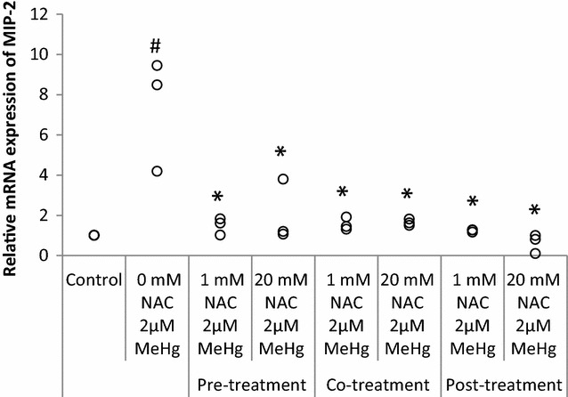
In a murine RAW264.7 macrophage cell line, MeHg-induced cytokine expressions were measured using real-time PCR. The suppressive effect of NAC was examined by putting it into the culture medium together with MeHg (co-treatment). In addition, pre- and post-treatment experiments were conducted, in which the cells were treated with NAC before and after MeHg exposure, respectively.
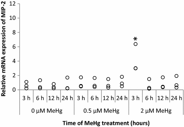
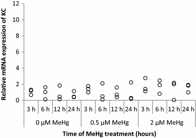
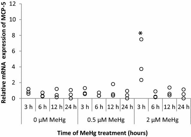
Exposure to a non-cytotoxic dose of MeHg up-regulated the mRNA expression of MIP-2 and MCP-5. On the other hand, KC expression was not induced in the presence of MeHg. Effect of MeHg on MIP-2 expressions was suppressed by pre-, co-, and post-treatment with NAC. However, the suppressive effect of pre-treatment was less than the post-treatment, which was as effective as co-treatment.
In functional homologues of human IL-8, only MIP-2 expression, not KC, was activated in the presence of non-cytotoxic dose of MeHg in murine RAW264.7 macrophage cell line. The more evident inhibitory effect of NAC observed in post-treatment experiments suggests a possible involvement of intracellular activities such as antioxidant effects.
本研究旨在检测在小鼠巨噬细胞中,非细胞毒性剂量的甲基汞(MeHg)存在时炎症细胞因子的表达情况,甲基汞被怀疑在MeHg暴露导致的脑损伤中起重要作用。我们重点研究了小鼠巨噬细胞炎性蛋白-2(MIP-2)、角质形成细胞趋化因子(KC)和单核细胞趋化蛋白-5(MCP-5)。MIP-2和KC分别是人类IL-8和MCP-5的小鼠功能同源物,MCP-5是人类MCP-1的小鼠功能同源物。此外,我们还检测了N-乙酰半胱氨酸(NAC)对MeHg诱导的炎症细胞因子的抑制作用。
在小鼠RAW264.7巨噬细胞系中,使用实时PCR检测MeHg诱导的细胞因子表达。通过将NAC与MeHg一起加入培养基(联合处理)来检测NAC的抑制作用。此外,还进行了预处理和后处理实验,即分别在MeHg暴露之前和之后用NAC处理细胞。
暴露于非细胞毒性剂量的MeHg可上调MIP-2和MCP-5的mRNA表达。另一方面,在MeHg存在的情况下未诱导KC表达。NAC的预处理、联合处理和后处理均可抑制MeHg对MIP-2表达的影响。然而,预处理的抑制作用小于后处理和联合处理,后处理与联合处理的效果相当。
在小鼠RAW264.7巨噬细胞系中,在非细胞毒性剂量的MeHg存在时,人类IL-8的功能同源物中只有MIP-2的表达被激活,而KC未被激活。在后处理实验中观察到的NAC更明显的抑制作用表明,细胞内活性如抗氧化作用可能参与其中。