Division of Signal Transduction and Growth Control, DKFZ/ZMBH Alliance, Heidelberg, Germany.
Functional Neuroanatomy, University of Heidelberg, Heidelberg, Germany.
BMC Cancer. 2018 Jan 30;18(1):103. doi: 10.1186/s12885-018-4007-4.
The poor prognosis for glioblastoma patients is caused by the diffuse infiltrative growth pattern of the tumor. Therefore, the molecular and cellular processes underlying cell migration continue to be a major focus of glioblastoma research. Emerging evidence supports the concept that the tumor microenvironment has a profound influence on the functional properties of tumor cells. Accordingly, substantial effort must be devoted to move from traditional two-dimensional migration assays to three-dimensional systems that more faithfully recapitulate the complex in vivo tumor microenvironment.
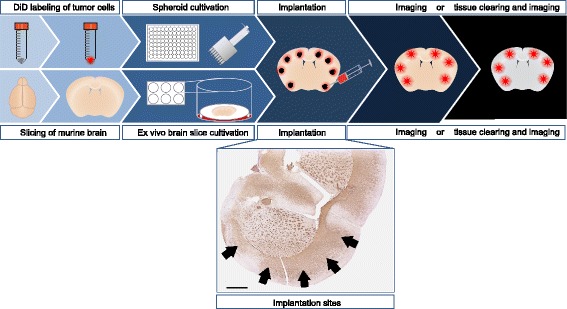
In order to mimic the tumor microenvironment of adult gliomas, we used adult organotypic brain slices as an invasion matrix for implanted, fluorescently labeled tumor spheroids. Cell invasion was imaged by confocal or epi-fluorescence microscopy and quantified by determining the average cumulative sprout length per spheroid. The tumor microenvironment was manipulated by treatment of the slice with small molecule inhibitors or using different genetically engineered mouse models as donors.
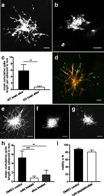
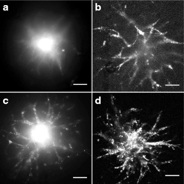
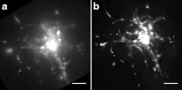
Both epi-fluorescence and confocal microscopy were applied to precisely quantify cell invasion in this ex vivo approach. Usage of a red-emitting membrane dye in addition to tissue clearing drastically improved epi-fluorescence imaging. Preparation of brain slices from of a genetically engineered mouse with a loss of a specific cell surface protein resulted in significantly impaired tumor cell invasion. Furthermore, jasplakinolide treatment of either tumor cells or brain slice significantly reduced tumor cell invasion.
We present an optimized invasion assay that closely reflects in vivo invasion by the implantation of glioma cells into organotypic adult brain slice cultures with a preserved cytoarchitecture. The diversity of applications including manipulation of the tumor cells as well as the microenvironment, permits the investigation of rate limiting factors of cell migration in a reliable context. This model will be a valuable tool for the discovery of the molecular mechanisms underlying glioma cell invasion and, ultimately, the development of novel therapeutic strategies.
胶质母细胞瘤患者的预后较差,是由于肿瘤的弥漫浸润性生长模式所致。因此,细胞迁移的分子和细胞过程仍然是胶质母细胞瘤研究的主要焦点。新出现的证据支持这样的概念,即肿瘤微环境对肿瘤细胞的功能特性有深远的影响。因此,必须大力将传统的二维迁移分析方法转变为更真实地再现复杂的体内肿瘤微环境的三维系统。
为了模拟成人胶质瘤的肿瘤微环境,我们使用成人器官型脑片作为植入的荧光标记肿瘤球体的侵袭基质。通过共聚焦或 epi 荧光显微镜对细胞侵袭进行成像,并通过确定每个球体的平均累积芽长来定量。通过对切片进行小分子抑制剂处理或使用不同的基因工程小鼠模型作为供体来操纵肿瘤微环境。
共聚焦和 epi 荧光显微镜都被应用于精确地定量这种离体方法中的细胞侵袭。除了组织清除外,使用红色发射膜染料极大地改善了 epi 荧光成像。从特定细胞表面蛋白缺失的基因工程小鼠制备脑片导致肿瘤细胞侵袭明显受损。此外,jasplakinolide 处理肿瘤细胞或脑片均显著降低了肿瘤细胞的侵袭。
我们提出了一种优化的侵袭分析方法,通过将胶质瘤细胞植入保留细胞结构的器官型成年脑片培养物中,紧密反映体内侵袭。包括对肿瘤细胞和微环境进行操作在内的多种应用,允许在可靠的背景下研究细胞迁移的限速因素。该模型将成为发现胶质母细胞瘤细胞侵袭的分子机制并最终开发新的治疗策略的有价值的工具。