Department of Radiology, Kyungpook National University Chilgok Hospital, Daegu, Republic of Korea.
Department of Radiology, School of Medicine, Kyungpook National University, Daegu, Republic of Korea.
Sci Rep. 2018 Feb 16;8(1):3181. doi: 10.1038/s41598-018-21554-z.
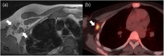
We aimed to investigate the value of breast magnetic resonance (MR) imaging and positron emission tomography-computed tomography (PET/CT) in predicting advanced axillary lymph node (ALN) metastases (ypN2-3) after neoadjuvant chemotherapy (NAC) in invasive ductal carcinoma patients. A total of 108 patients with invasive ductal carcinoma underwent breast MR imaging and PET/CT both before and after NAC (termed initial staging and restaging, respectively). The number of positive ALNs and the short diameter (SD) of the largest ALN on breast MR imaging and maximal standardized uptake value (SUVmax) in the ALNs on PET/CT were evaluated. Odds ratio (OR) for prediction of advanced ALN metastases was calculated. The negative predictive value (NPV) of restaging imaging for exclusion of advanced ALN metastases was also calculated. Patients with advanced ALN metastases were more likely to have a higher number (≥2) of positive LNs (OR, 8.06; P = 0.015) on restaging MR imaging. No clinico-pathological factors were significantly associated with advanced ALN metastases. With restaging MR imaging, PET/CT, and MR imaging plus PET/CT, the NPV for excluding advanced ALN metastases was 97.3%, 94.4%, and 100.0%. A higher number of positive ALNs on restaging MR imaging was an independent predictor for advanced ALN metastases after NAC.
我们旨在探讨乳腺磁共振(MR)成像和正电子发射断层扫描-计算机断层扫描(PET/CT)在预测新辅助化疗(NAC)后浸润性导管癌患者腋窝淋巴结(ALN)转移(ypN2-3)中的价值。共有 108 例浸润性导管癌患者分别在 NAC 前(初始分期)和后(重新分期)进行了乳腺 MR 成像和 PET/CT 检查。评估了乳腺 MR 成像上阳性 ALN 的数量和最大 ALN 的短直径(SD)以及 PET/CT 上 ALN 的最大标准化摄取值(SUVmax)。计算了预测高级 ALN 转移的优势比(OR)。还计算了重新分期成像排除高级 ALN 转移的阴性预测值(NPV)。具有高级 ALN 转移的患者更有可能在重新分期的 MR 成像上具有更多的阳性 LNs(OR,8.06;P=0.015)。没有临床病理因素与高级 ALN 转移明显相关。对于重新分期的 MR 成像、PET/CT 和 MR 成像加 PET/CT,排除高级 ALN 转移的 NPV 分别为 97.3%、94.4%和 100.0%。重新分期的 MR 成像上阳性 ALN 数量较多是 NAC 后高级 ALN 转移的独立预测因素。