Department of Nuclear Medicine, Huadong Hospital, Fudan University, Shanghai, China (mainland).
Med Sci Monit. 2018 Mar 6;24:1373-1378. doi: 10.12659/msm.905664.
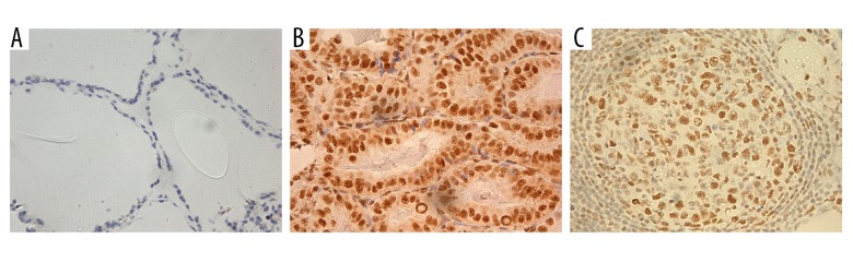
BACKGROUND This paper aimed to evaluate the expression of REGg and characterize its clinical significance in papillary thyroid carcinoma (PTC). MATERIAL AND METHODS In total, 54 patients with PTC who underwent partial or total thyroidectomy and cervical node dissection for PTC from February 2009 to September 2011 were retrospectively reviewed. Thyroid specimens and metastatic lymph nodes from 54 patients and normal thyroid tissues obtained from 13 volunteers were collected and analyzed. Tumor size, T-stage, and lymph nodes metastasis were recorded based on surgical pathology. Immunohistochemical (IHC) technology was performed to analyze REGg protein expression level. Corrections between the expression of REGγ and the clinicopathological factors were analyzed. RESULTS All the normal thyroid tissues were REGg-negative. REGγ was positive in 75.9% (41/54) of PTC tissues, of which 29 cases (29/42, 69.0%) were in T1-T2 stage and 12 cases (12/12,100%) were in T3-T4 stage. Positive REGγ was found in 21 cases (21/24, 87.5%) in T1-T2 stage with lymph nodes metastasis, while 11 cases were in T3-T4 stage with metastases to lymph nodes (11/11, 100%). High level of REGγ expression was significantly correlated with T-stage (P<0.05) and lymph node metastases (P<0.05). In addition, there was no statistically significant difference between the expression of REGγ and age, sex, tumor size, or tumor multiplicity (P>0.05). Using binary logistic regression model, positive REGγ was identified as a significant independent predictor factor of lymph node metastasis in PTC. CONCLUSIONS High expression of REGg seemed positively correlated with T-stage and lymph node metastasis in PTC tissues.
背景 本研究旨在评估 REGg 的表达并探讨其在甲状腺乳头状癌(PTC)中的临床意义。
材料与方法 回顾性分析 2009 年 2 月至 2011 年 9 月因 PTC 接受甲状腺部分或全切除术和颈部淋巴结清扫术的 54 例患者的临床病理资料。收集并分析 54 例患者的甲状腺标本及转移淋巴结和 13 例志愿者的正常甲状腺组织。根据手术病理记录肿瘤大小、T 分期和淋巴结转移情况。采用免疫组织化学(IHC)技术分析 REGγ 蛋白表达水平。分析 REGγ 表达与临床病理因素的相关性。
结果 所有正常甲状腺组织均为 REGγ 阴性。在 54 例 PTC 组织中,REGγ 阳性率为 75.9%(41/54),其中 T1-T2 期 29 例(29/42,69.0%),T3-T4 期 12 例(12/12,100%)。在 T1-T2 期有淋巴结转移的 24 例中,21 例(21/24,87.5%)REGγ 阳性,而在 T3-T4 期有淋巴结转移的 11 例中,11 例(11/11,100%)REGγ 阳性。REGγ 高表达与 T 分期(P<0.05)和淋巴结转移(P<0.05)显著相关。此外,REGγ 的表达与年龄、性别、肿瘤大小或肿瘤数量无关(P>0.05)。采用二元逻辑回归模型,REGγ 阳性被确定为 PTC 淋巴结转移的独立预测因子。
结论 在 PTC 组织中,REGγ 的高表达似乎与 T 分期和淋巴结转移呈正相关。