Eye Center, Medical Center, Medical Faculty, University of Freiburg, Freiburg, Germany.
PLoS One. 2018 Mar 12;13(3):e0191338. doi: 10.1371/journal.pone.0191338. eCollection 2018.
Retinal vein occlusion (RVO) has been investigated in several laser-induced animal models using pigs, rabbits and rats. However, laser-induced RVO has been rarely reported in mice, despite the impressive number of available mutants, ease of handling and cost effectiveness. The aim of this study was to further assess the feasibility of a RVO mouse model for gene expression analysis and its possible use to investigate effects of hypoxia.
C57Bl/6J mice were injected with eosin Y for photo-sensitization. Subsequently, large retinal veins were laser-treated in one eye to induce vascular occlusion. Contralateral control eyes received non-occlusive retinal laser treatment sparing large vessels. The animals were followed for up to eight days and assessed by funduscopy, angiography, hypoxyprobe staining, histopathology and gene expression analysis by qPCR and RNA sequencing (RNAseq). Another group of mice was left untreated and studied at a single time point to determine baseline characteristics.
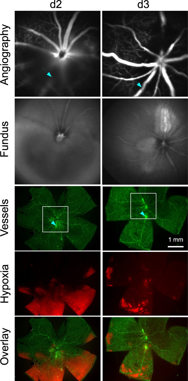
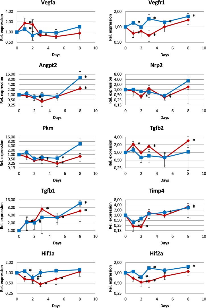
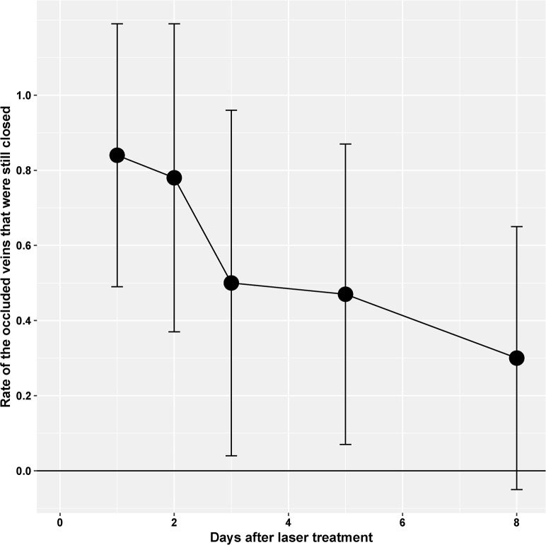
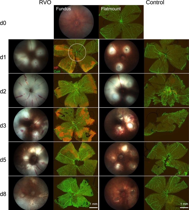
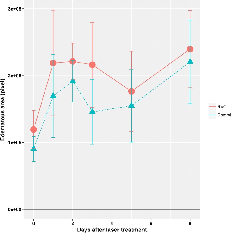
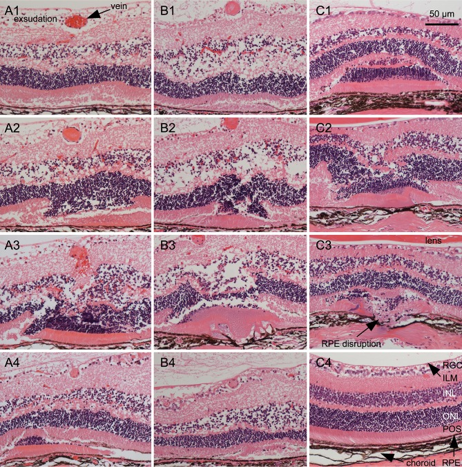
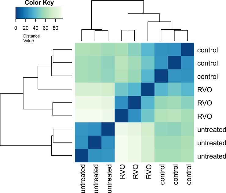
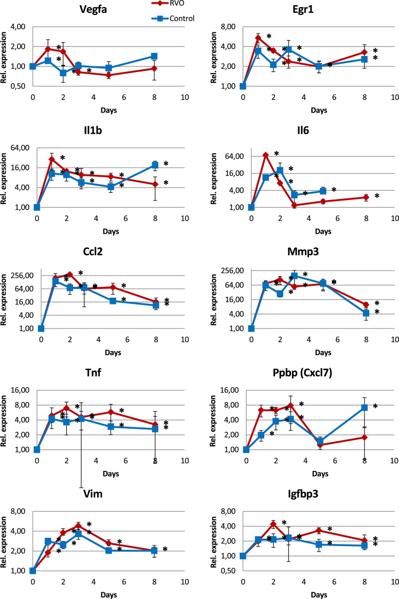
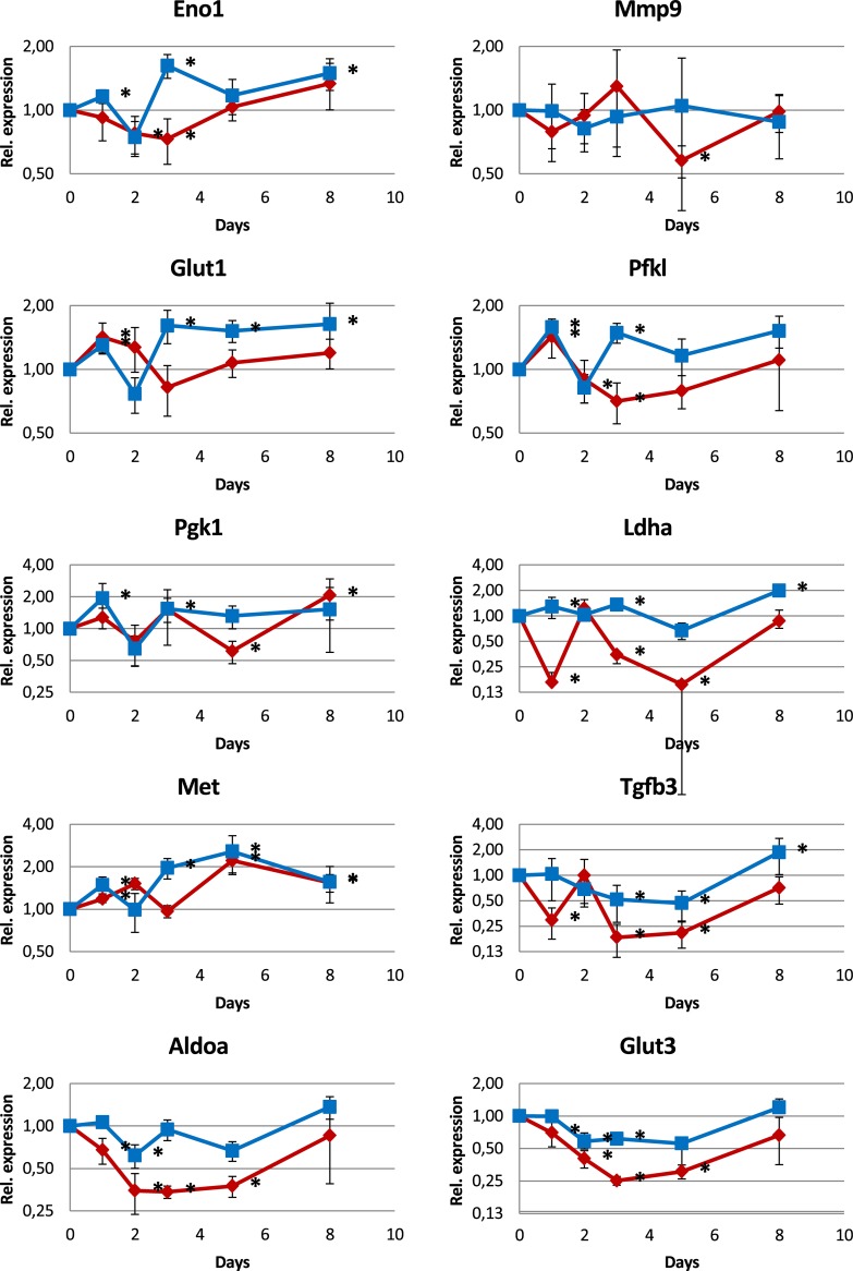
Laser-induced RVO persisted in half of the treated veins for three days, and in a third of the veins for the whole observation period of 8 days. Funduscopy revealed large areas of retinal swelling in all laser-treated eyes, irrespective of vascular targeting or occlusion status. Damage of the outer retina, retinal pigment epithelium (RPE), and even choroid and sclera at the laser site was observed in histological sections. Genes associated with inflammation or cell damage were highly up-regulated in all laser-treated eyes as detected by RNAseq and qPCR. Retinal hypoxia was observed by hypoxyprobe staining in all RVO eyes for up to 5 days with a maximal extension at days 2 and 3, but no significant RVO-dependent changes in gene expression were detected for angiogenesis- or hypoxia-related genes.
The laser-induced RVO mouse model is characterized by a predominant general inflammatory and tissue damage response, which may obscure distinct hypoxia- and angiogenesis-related effects. A non-occlusive laser treatment control is essential to allow for proper data interpretation and should be mandatory in animal studies of laser-induced RVO to dissect laser-induced tissue damage from vascular occlusion effects.
视网膜静脉阻塞(RVO)已在几种使用猪、兔和大鼠的激光诱导动物模型中进行了研究。然而,尽管有大量可用的突变体、易于处理和成本效益,激光诱导的 RVO 在小鼠中很少有报道。本研究旨在进一步评估 RVO 小鼠模型用于基因表达分析的可行性,及其用于研究缺氧影响的可能用途。
C57Bl/6J 小鼠用曙红 Y 进行光致敏。随后,在一只眼中对大的视网膜静脉进行激光处理以诱导血管闭塞。对侧对照眼接受非闭塞性视网膜激光治疗,不损伤大血管。对动物进行长达 8 天的随访,并通过眼底检查、血管造影、缺氧探针染色、组织病理学和 qPCR 和 RNA 测序(RNAseq)进行基因表达分析。另一组小鼠未进行治疗,并在一个时间点进行研究以确定基线特征。
激光诱导的 RVO 在处理的静脉中有一半持续了 3 天,在三分之一的静脉中持续了整个 8 天的观察期。眼底检查显示所有激光治疗眼均出现大面积视网膜肿胀,无论血管靶向或闭塞状态如何。在组织学切片中观察到外视网膜、视网膜色素上皮(RPE)甚至脉络膜和巩膜在激光部位的损伤。RNAseq 和 qPCR 检测到,所有激光治疗眼的炎症或细胞损伤相关基因均高度上调。缺氧探针染色显示,所有 RVO 眼中的视网膜缺氧持续至 5 天,在第 2 和第 3 天达到最大扩展,但未检测到血管生成或缺氧相关基因的明显 RVO 依赖性变化。
激光诱导的 RVO 小鼠模型的特征是主要的炎症和组织损伤反应,这可能掩盖了明显的缺氧和血管生成相关效应。非闭塞性激光治疗对照对于适当的数据解释至关重要,并且在激光诱导的 RVO 动物研究中应是强制性的,以将激光诱导的组织损伤与血管闭塞效应分开。