Division of Cardiology, Labatt Family Heart Center, Toronto, Ontario, Canada.
Translational Medicine, Hospital for Sick Children and University of Toronto, Ontario, Canada.
J Am Heart Assoc. 2018 Mar 29;7(7):e007928. doi: 10.1161/JAHA.117.007928.
Development of right ventricular (RV) hypertension eventually contributes to RV and left ventricular (LV) myocardial fibrosis and dysfunction. The molecular mechanisms are not fully elucidated.
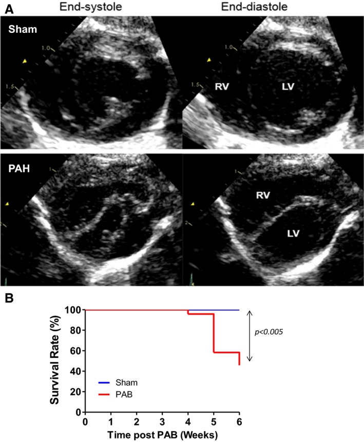
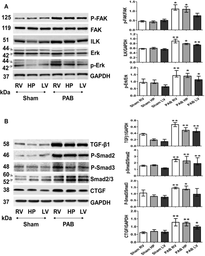
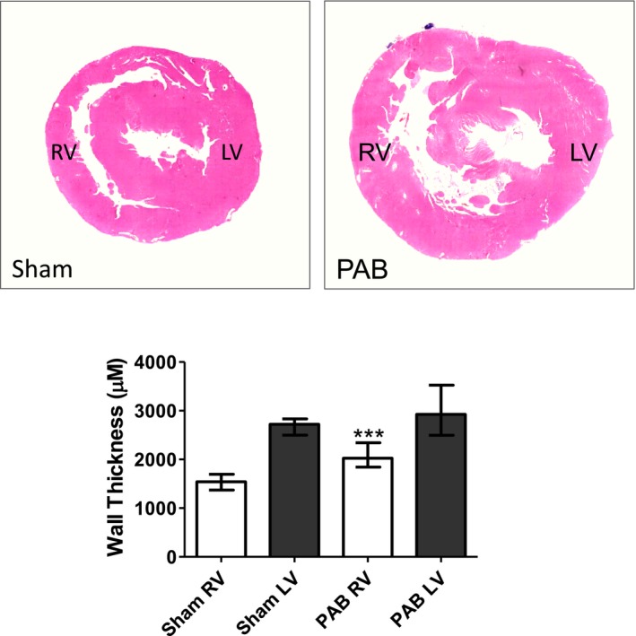
Pulmonary artery banding was used to induce RV hypertension in rats in vivo. Then, we evaluated cardiac function and regional remodeling 6 weeks after pulmonary artery banding. To further elucidate mechanisms responsible for regional cardiac remodeling, we also mimicked RV hypertensive stress by cyclic mechanical stretching applied to confluent cultures of cardiac fibroblasts, isolated from the RV free wall, septal hinge points, and LV free wall. Echocardiography and catheter evaluation demonstrated that rats in the pulmonary artery banding group developed RV hypertension with leftward septal displacement, LV compression, and increased LV end-diastolic pressures. Picrosirius red staining indicated that pulmonary artery banding induced marked RV fibrosis and dysfunction, with prominent fibrosis and elastin deposition at the septal hinge points but less LV fibrosis. These changes were associated with proportionally increased expressions of integrin-β1 and profibrotic signaling proteins, including phosphorylated Smad2/3 and transforming growth factor-β1. Moreover, mechanically stretched fibroblasts also expressed significantly increased levels of α-smooth muscle actin, integrin-β1, transforming growth factor-β1, collagen I deposition, and wrinkle formation on gel assays, consistent with myofibroblast transformation. These changes were not observed in parallel cultures of mechanically stretched fibroblasts, preincubated with the integrin inhibitor (BTT-3033).
Experimentally induced RV hypertension triggers regional RV, hinge-point, and LV integrin β1-dependent mechanotransduction signaling pathways that eventually trigger myocardial fibrosis via transforming growth factor-β1 signaling. Reduced LV fibrosis and preserved global function, despite geometrical and pressure aberrations, suggest a possible elastin-mediated protective mechanism at the septal hinge points.
右心室(RV)高血压的发展最终会导致 RV 和左心室(LV)心肌纤维化和功能障碍。其分子机制尚未完全阐明。
在体内使用肺动脉缩窄术诱导大鼠 RV 高血压。然后,我们评估了肺动脉缩窄 6 周后的心脏功能和区域性重塑。为了进一步阐明导致区域性心脏重塑的机制,我们还通过对来源于 RV 游离壁、室间隔铰链点和 LV 游离壁的心脏成纤维细胞的汇合培养物施加周期性机械拉伸来模拟 RV 高血压应激。超声心动图和导管评估表明,肺动脉缩窄组大鼠出现 RV 高血压,伴有左室间隔移位、LV 受压和 LV 舒张末期压升高。苦味酸红染色表明肺动脉缩窄诱导明显的 RV 纤维化和功能障碍,在室间隔铰链点出现明显的纤维化和弹性蛋白沉积,但 LV 纤维化较少。这些变化与整合素-β1 和促纤维化信号蛋白(包括磷酸化 Smad2/3 和转化生长因子-β1)的比例增加有关。此外,机械拉伸的成纤维细胞在凝胶试验中也表现出明显增加的α-平滑肌肌动蛋白、整合素-β1、转化生长因子-β1、胶原 I 沉积和皱纹形成,与肌成纤维细胞转化一致。这些变化在预先用整合素抑制剂(BTT-3033)孵育的机械拉伸成纤维细胞的平行培养物中没有观察到。
实验诱导的 RV 高血压引发了 RV、铰链点和 LV 整合素β1 依赖性机械转导信号通路,最终通过转化生长因子-β1 信号触发心肌纤维化。尽管存在几何和压力异常,但 LV 纤维化减少和整体功能保留表明,在室间隔铰链点可能存在弹性蛋白介导的保护机制。