• 文献检索
  • 文档翻译
  • 深度研究
  • 学术资讯
  • Suppr Zotero 插件Zotero 插件
  • 邀请有礼
  • 套餐&价格
  • 历史记录
应用&插件
Suppr Zotero 插件Zotero 插件浏览器插件Mac 客户端Windows 客户端微信小程序
定价
高级版会员购买积分包购买API积分包
服务
文献检索文档翻译深度研究API 文档MCP 服务
关于我们
关于 Suppr公司介绍联系我们用户协议隐私条款
关注我们

Suppr 超能文献

核心技术专利:CN118964589B侵权必究
粤ICP备2023148730 号-1Suppr @ 2026

文献检索

告别复杂PubMed语法,用中文像聊天一样搜索,搜遍4000万医学文献。AI智能推荐,让科研检索更轻松。

立即免费搜索

文件翻译

保留排版,准确专业,支持PDF/Word/PPT等文件格式,支持 12+语言互译。

免费翻译文档

深度研究

AI帮你快速写综述,25分钟生成高质量综述,智能提取关键信息,辅助科研写作。

立即免费体验

脑膜瘤质地:磁共振成像特征、手术所见与组织病理学特征之间的相关性

Meningioma Consistency: Correlation Between Magnetic Resonance Imaging Characteristics, Operative Findings, and Histopathological Features.

作者信息

Alyamany Mahmoud, Alshardan Mohammad M, Jamea Abdullah Abu, ElBakry Nahid, Soualmi Lahbib, Orz Yasser

机构信息

Department of Neurosurgery, National Neuroscience Institute, King Saud Bin Abdulaziz University for Health Sciences, King Fahad Medical City, Riyadh, Saudi Arabia.

Neurosurgery Division, University of Ottawa, Ottawa, Ontario, Canada.

出版信息

Asian J Neurosurg. 2018 Apr-Jun;13(2):324-328. doi: 10.4103/1793-5482.228515.

DOI:10.4103/1793-5482.228515
PMID:29682029
原文链接:https://pmc.ncbi.nlm.nih.gov/articles/PMC5898100/
Abstract

INTRODUCTION

Intracranial meningiomas account for 30% of all primary intracranial tumors. Surgical resection remains the mainstay of the treatment for meningiomas. The magnetic resonance of intracranial meningiomas has been largely discussed in many reports of the radiological and neurosurgical literature. To date, a few studies have been attempted to differentiate the tumor characteristics of meningiomas based on magnetic resonance imaging (MRI) studies.

OBJECTIVE

The objective of the study is to evaluate the relationship between MRI signal characteristics of intracranial meningiomas and consistency of tumor using objective measures.

MATERIALS AND METHODS

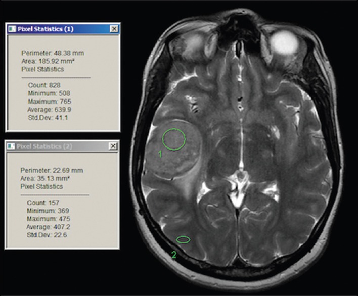
A prospective study included all the patients who were admitted for surgery with an MRI finding suggestive of meningioma. All patients were subjected to routine radiological investigations. Surgical resection was performed for patients eligible for surgery using cavitron ultrasonic aspirator (CUSA). The relationship and correlation between the radiological, intraoperative measurements and the histopathological diagnosis were studied. The tumor consistency was measured using mean CUSA level. Intensity on T2, fluid-attenuated inversion recovery (FLAIR), and diffusion-weighted imaging (DWI) was measured using circular regions of interest (ROI) on the MRI. Multiple ROIs were placed initially on the lesions avoiding the obvious blood vessels, if any, then on the brain cortex to avoid the vasogenic edema. The mean ROI (mROI) results from the lesion were subtracted from the mean ROI from the brain cortex for each lesion to achieve normalized ratio. The results of lesion mROI-cortex mROI were compared to the operative and histopathology results using Pearson's correlation test and linear regression test.

RESULTS

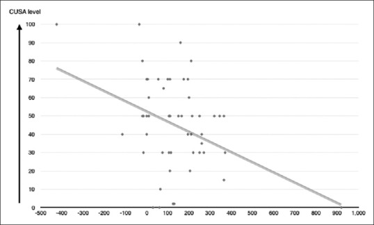
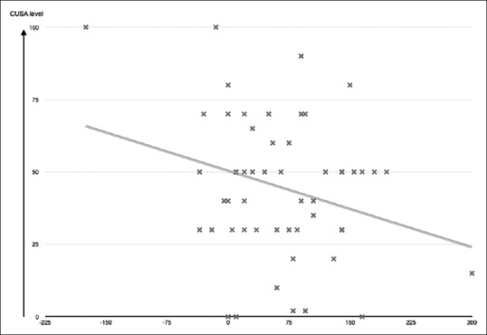
The total number of patients was seventy. The mean age of the patients was 51 ± 14.8, with 72% of them being females and 28% males. There was a strong statistically significant ( = 0.046) and ( = 0.003) correlation between mean CUSA and FLAIR mROI difference or T2 mROI difference, respectively. On the other hand, there was an inversely proportional relationship between mean CUSA and FLAIR mROI difference and mean CUSA and T2 mROI difference. The value of the regression test () shows that there was a slight linear relationship between FLAIR mROI difference or T2 mROI difference and mean CUSA values, in which the mean CUSA value = 50.1 + (-0.088) × FLAIR mROI difference ( = -0.273, = 0.046) or mean CUSA value = 50.8 + (-0.055) × T2 mROI difference ( = 0.4, = 0.003). There was no statistical significance in the relation between CUSA values and tumor histological subtypes, DWI values, age, or gender.

CONCLUSION

This study presents a new objective method to measure the consistency of intracranial meningiomas based on a simple algorithmic formula. Such information will aid in planning surgery and assessing the resectability of the tumor. To date, this is the first objective measurement of meningioma consistency based on MRI studies and objective intraoperative evaluation.

https://cdn.ncbi.nlm.nih.gov/pmc/blobs/358b/5898100/49520590bef4/AJNS-13-324-g004.jpg
https://cdn.ncbi.nlm.nih.gov/pmc/blobs/358b/5898100/3723af45f5fe/AJNS-13-324-g001.jpg
https://cdn.ncbi.nlm.nih.gov/pmc/blobs/358b/5898100/a2019ec7c11a/AJNS-13-324-g003.jpg
https://cdn.ncbi.nlm.nih.gov/pmc/blobs/358b/5898100/49520590bef4/AJNS-13-324-g004.jpg
https://cdn.ncbi.nlm.nih.gov/pmc/blobs/358b/5898100/3723af45f5fe/AJNS-13-324-g001.jpg
https://cdn.ncbi.nlm.nih.gov/pmc/blobs/358b/5898100/a2019ec7c11a/AJNS-13-324-g003.jpg
https://cdn.ncbi.nlm.nih.gov/pmc/blobs/358b/5898100/49520590bef4/AJNS-13-324-g004.jpg
摘要

引言

颅内脑膜瘤占所有原发性颅内肿瘤的30%。手术切除仍然是脑膜瘤治疗的主要手段。颅内脑膜瘤的磁共振成像在许多放射学和神经外科学文献报告中已有大量讨论。迄今为止,已有一些研究试图基于磁共振成像(MRI)研究来区分脑膜瘤的肿瘤特征。

目的

本研究的目的是使用客观测量方法评估颅内脑膜瘤的MRI信号特征与肿瘤质地之间的关系。

材料与方法

一项前瞻性研究纳入了所有因MRI检查发现提示脑膜瘤而入院接受手术的患者。所有患者均接受常规放射学检查。对于符合手术条件的患者,使用超声吸引器(CUSA)进行手术切除。研究了放射学、术中测量结果与组织病理学诊断之间的关系及相关性。使用CUSA平均水平测量肿瘤质地。在MRI上,使用圆形感兴趣区(ROI)测量T2加权像、液体衰减反转恢复序列(FLAIR)及扩散加权成像(DWI)上的信号强度。最初在病变上放置多个ROI,避开明显血管(如有),然后在脑皮质上放置ROI以避免血管源性水肿。将每个病变的病变平均ROI(mROI)结果减去脑皮质的平均ROI,以获得标准化比值。使用Pearson相关检验和线性回归检验,将病变mROI-皮质mROI的结果与手术及组织病理学结果进行比较。

结果

患者总数为70例。患者的平均年龄为51±14.8岁,其中72%为女性,28%为男性。平均CUSA与FLAIR mROI差值或T2 mROI差值之间分别存在高度统计学显著相关性(P = 0.046)和(P = 0.003)。另一方面,平均CUSA与FLAIR mROI差值以及平均CUSA与T2 mROI差值之间呈反比关系。回归检验的值(R)表明,FLAIR mROI差值或T2 mROI差值与平均CUSA值之间存在轻微线性关系,其中平均CUSA值 = 50.1 +(-0.088)×FLAIR mROI差值(R = -0.273,P = 0.046)或平均CUSA值 = 50.8 +(-0.055)×T2 mROI差值(R = 0.4,P = 0.003)。CUSA值与肿瘤组织学亚型、DWI值、年龄或性别之间的关系无统计学意义。

结论

本研究提出了一种基于简单算法公式测量颅内脑膜瘤质地的新客观方法。此类信息将有助于手术规划及评估肿瘤的可切除性。迄今为止,这是基于MRI研究和客观术中评估对脑膜瘤质地进行的首次客观测量。

相似文献

1
Meningioma Consistency: Correlation Between Magnetic Resonance Imaging Characteristics, Operative Findings, and Histopathological Features.脑膜瘤质地:磁共振成像特征、手术所见与组织病理学特征之间的相关性
Asian J Neurosurg. 2018 Apr-Jun;13(2):324-328. doi: 10.4103/1793-5482.228515.
2
Meningioma consistency prediction utilizing tumor to cerebellar peduncle intensity on T2-weighted magnetic resonance imaging sequences: TCTI ratio.利用 T2 加权磁共振成像序列中肿瘤与小脑脑桥强度比预测脑膜瘤质地:TCTI 比值。
J Neurosurg. 2017 Jan;126(1):242-248. doi: 10.3171/2016.1.JNS152329. Epub 2016 Apr 8.
3
The Predictive Value of Conventional Magnetic Resonance Imaging Sequences on Operative Findings and Histopathology of Intracranial Meningiomas: A Prospective Study.常规磁共振成像序列对颅内脑膜瘤手术发现和组织病理学的预测价值:一项前瞻性研究。
Neurol India. 2019 Nov-Dec;67(6):1439-1445. doi: 10.4103/0028-3886.273632.
4
Supratentorial Meningioma Consistency Prediction Utilizing Tumor to Cerebellar Peduncle Intensity on T1 and T2-Weighted and Fluid Attenuated Inversion Recovery Magnetic Resonance Imaging Sequences.利用 T1、T2 加权和液体衰减反转恢复磁共振成像序列上肿瘤与小脑脑桥脚强度预测幕上脑膜瘤硬度。
World Neurosurg. 2023 Feb;170:e180-e187. doi: 10.1016/j.wneu.2022.10.097. Epub 2022 Nov 1.
5
Benefits of Combined MRI Sequences in Meningioma Consistency Prediction: A Prospective Study of 287 Consecutive Patients.联合MRI序列在脑膜瘤质地预测中的益处:对287例连续患者的前瞻性研究
Asian J Neurosurg. 2022 Dec 10;17(4):614-620. doi: 10.1055/s-0042-1758849. eCollection 2022 Dec.
6
Prediction of hard meningiomas: quantitative evaluation based on the magnetic resonance signal intensity.硬脑膜瘤的预测:基于磁共振信号强度的定量评估
Acta Radiol. 2016 Mar;57(3):333-40. doi: 10.1177/0284185115578323. Epub 2015 Mar 29.
7
Diffusion tensor magnetic resonance imaging for predicting the consistency of intracranial meningiomas.用于预测颅内脑膜瘤一致性的扩散张量磁共振成像
Acta Neurochir (Wien). 2014 Oct;156(10):1837-45. doi: 10.1007/s00701-014-2149-y. Epub 2014 Jul 8.
8
Predicting Consistency of Meningioma by Magnetic Resonance Imaging.通过磁共振成像预测脑膜瘤的一致性
J Neurol Surg B Skull Base. 2015 Jun;76(3):225-9. doi: 10.1055/s-0034-1543965. Epub 2015 Jan 21.
9
Multi-parametric qualitative and quantitative MRI assessment as predictor of histological grading in previously treated meningiomas.多参数定性和定量 MRI 评估可预测既往治疗脑膜瘤的组织学分级。
Neuroradiology. 2020 Nov;62(11):1441-1449. doi: 10.1007/s00234-020-02476-y. Epub 2020 Jun 24.
10
Application of magnetic resonance fingerprinting to differentiate grade I transitional and fibrous meningiomas from meningothelial meningiomas.磁共振指纹识别技术在鉴别I级过渡型和纤维型脑膜瘤与脑膜皮型脑膜瘤中的应用。
Quant Imaging Med Surg. 2021 Apr;11(4):1447-1457. doi: 10.21037/qims-20-732.

引用本文的文献

1
Predicting intraoperative meningioma consistency using features from standard MRI sequences: a preoperative evaluation.利用标准MRI序列特征预测术中脑膜瘤质地:一项术前评估
Acta Neurochir (Wien). 2025 Jun 21;167(1):173. doi: 10.1007/s00701-025-06582-9.
2
Stiffness analysis of meningiomas using neural network-based inversion on MR Elastography.基于神经网络反演的磁共振弹性成像对脑膜瘤的硬度分析
Annu Int Conf IEEE Eng Med Biol Soc. 2024 Jul;2024:1-5. doi: 10.1109/EMBC53108.2024.10781605.
3
Histogram analysis of mono-exponential, bi-exponential and stretched-exponential diffusion-weighted MR imaging in predicting consistency of meningiomas.

本文引用的文献

1
Diffusion tensor magnetic resonance imaging for predicting the consistency of intracranial meningiomas.用于预测颅内脑膜瘤一致性的扩散张量磁共振成像
Acta Neurochir (Wien). 2014 Oct;156(10):1837-45. doi: 10.1007/s00701-014-2149-y. Epub 2014 Jul 8.
2
Diagnostic value of apparent diffusion coefficient for the accurate assessment and differentiation of intracranial meningiomas.表观扩散系数对颅内脑膜瘤准确评估及鉴别诊断的价值
Acta Radiol Short Rep. 2013 Nov 23;2(7):2047981613512484. doi: 10.1177/2047981613512484. eCollection 2013.
3
CBTRUS statistical report: primary brain and central nervous system tumors diagnosed in the United States in 2005-2009.
单指数、双指数和拉伸指数扩散加权磁共振成像直方图分析在预测脑膜瘤一致性中的应用。
Cancer Imaging. 2023 Dec 5;23(1):117. doi: 10.1186/s40644-023-00633-z.
4
Utility of texture analysis for objective quantitative ex vivo assessment of meningioma consistency: method proposal and validation.纹理分析在脑膜瘤硬度的客观定量体外评估中的应用:方法的提出与验证。
Acta Neurochir (Wien). 2023 Dec;165(12):4203-4211. doi: 10.1007/s00701-023-05867-1. Epub 2023 Dec 4.
5
Relationship Between Quantitative Tumor Consistency and Pathological Factors in Intracranial Meningioma.颅内脑膜瘤肿瘤一致性定量与病理因素的关系。
Acta Neurochir (Wien). 2023 Oct;165(10):2895-2902. doi: 10.1007/s00701-023-05712-5. Epub 2023 Jul 11.
6
Differentiation between various types and subtypes of intracranial meningiomas with advanced MRI.利用先进的磁共振成像技术鉴别颅内脑膜瘤的各种类型和亚型。
SA J Radiol. 2022 Oct 26;26(1):2480. doi: 10.4102/sajr.v26i1.2480. eCollection 2022.
7
Isolated incomplete third cranial nerve palsy due to presumed cavernous sinus meningioma.因推测为海绵窦脑膜瘤导致的孤立性不完全性动眼神经麻痹。
Oman J Ophthalmol. 2022 Mar 2;15(1):104-106. doi: 10.4103/ojo.ojo_406_20. eCollection 2022 Jan-Apr.
8
Can an Imaging Marker of Consistency Predict Intraoperative Experience and Clinical Outcomes for Vestibular Schwannomas? A Retrospective Review.一致性成像标志物能否预测前庭神经鞘瘤的术中体验和临床结果?一项回顾性研究。
J Neurol Surg B Skull Base. 2021 Apr;82(2):251-257. doi: 10.1055/s-0039-1697026. Epub 2019 Sep 24.
CBTRUS统计报告:2005 - 2009年在美国诊断出的原发性脑和中枢神经系统肿瘤
Neuro Oncol. 2012 Nov;14 Suppl 5(Suppl 5):v1-49. doi: 10.1093/neuonc/nos218.
4
Predictors of meningioma consistency: A study in 243 consecutive cases.脑膜瘤硬度的预测因素:243 例连续病例研究。
Acta Neurochir (Wien). 2012 Aug;154(8):1383-9. doi: 10.1007/s00701-012-1427-9. Epub 2012 Jun 29.
5
Use of preoperative magnetic resonance imaging T1 and T2 sequences to determine intraoperative meningioma consistency.使用术前磁共振成像T1和T2序列来确定术中脑膜瘤的质地。
Surg Neurol Int. 2011;2:142. doi: 10.4103/2152-7806.85983. Epub 2011 Oct 12.
6
Differentiation of fibroblastic meningiomas from other benign subtypes using diffusion tensor imaging.使用扩散张量成像技术鉴别纤维母细胞型脑膜瘤与其他良性亚型。
J Magn Reson Imaging. 2007 Apr;25(4):703-8. doi: 10.1002/jmri.20887.
7
Low-field MR imaging of meningiomas including dynamic contrast enhancement study: evaluation of surgical and histopathologic characteristics.脑膜瘤的低场磁共振成像,包括动态对比增强研究:手术及组织病理学特征评估
AJNR Am J Neuroradiol. 2006 Nov-Dec;27(10):2128-34.
8
Clinical and radiological features related to the growth potential of meningioma.与脑膜瘤生长潜能相关的临床和影像学特征
Neurosurg Rev. 2006 Oct;29(4):293-6; discussion 296-7. doi: 10.1007/s10143-006-0039-3. Epub 2006 Sep 5.
9
Comparison between neuroimaging classifications and histopathological diagnoses using an international multicenter brain tumor magnetic resonance imaging database.利用国际多中心脑肿瘤磁共振成像数据库对神经影像学分类与组织病理学诊断进行比较。
J Neurosurg. 2006 Jul;105(1):6-14. doi: 10.3171/jns.2006.105.1.6.
10
Epidemiology of intracranial meningioma.颅内脑膜瘤的流行病学
Neurosurgery. 2005 Dec;57(6):1088-95; discussion 1088-95. doi: 10.1227/01.neu.0000188281.91351.b9.