Cao Chenxi, Cao Zhengming, Liu Guangyu, Liu Songyang, Ye Yanqi, Sun Tiezheng
Arthritis Clinic and Research Center, Peking University, People's Hospital, Beijing, 100044, People's Republic of China.
BMC Musculoskelet Disord. 2018 May 24;19(1):163. doi: 10.1186/s12891-018-2087-6.
Angioleiomyoma is a very rare benign solitary soft tissue neoplasm originating from smooth muscle layer of blood vessels. The tumor is usually located in the subcutis or the superficial fasciae, but less often in the deep fasciae, especially rare in the knee joint cavity. Diagnosis is frequently delayed or misdiagnosed as loose body or anterior knee pain because of its rare occurrence and poor awareness of physicians. Few studies have presented intra-articular angioleiomyoma and such cases become rarer and more difficult to diagnose when it presents as loose body.
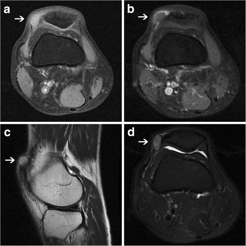
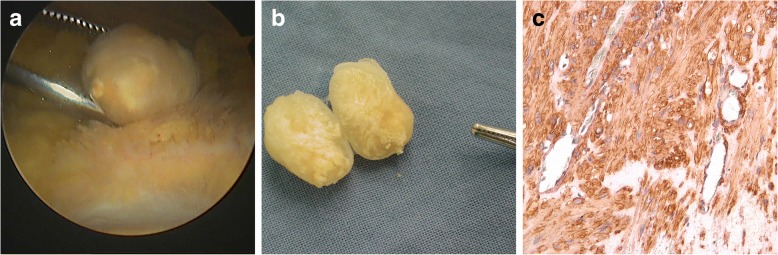
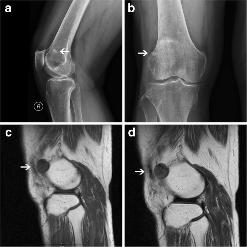
Two patients, a middle-aged man and an old woman, presented to our outpatient clinic with persistent anterior knee pain and both of them suffered from a solitary mass in the right knee that had slowly enlarged. One of two patients showed negative in the routine radiographic imaging and the other showed a "loose body" beside the lateral femoral condyle in the knee. MRI showed both a well-demarcated intra-articular mass of isointense signal to muscle on T1-weighted images and heterogeneous intensity on T2-weighted images. Their tumors were excised under arthroscopy finally, with the pathological results revealed vascular leiomyomas. They both recovered well with pain free after operation and no signs of recurrence were seen at the 7-year follow-up.
This case report illustrates the atypical locations of angioleiomyoma in the knee joint should arouse our attention and be included in the differential diagnosis of nodular lesions mimicking loose bodies.
血管平滑肌瘤是一种非常罕见的良性孤立性软组织肿瘤,起源于血管的平滑肌层。该肿瘤通常位于皮下或浅筋膜,但较少位于深筋膜,在膝关节腔内尤为罕见。由于其罕见性以及医生对其认识不足,诊断常常延迟或被误诊为游离体或膝关节前疼痛。很少有研究报道关节内血管平滑肌瘤,当它表现为游离体时,此类病例变得更加罕见且难以诊断。
两名患者,一名中年男性和一名老年女性,因持续性膝关节前疼痛前来我院门诊就诊,两人右膝均有一个缓慢增大的孤立肿块。两名患者中的一名在常规影像学检查中结果为阴性,另一名在膝关节外侧髁旁显示有一个“游离体”。磁共振成像(MRI)显示,在T1加权图像上,关节内肿块边界清晰,信号强度与肌肉等信号,在T2加权图像上信号强度不均匀。他们的肿瘤最终在关节镜下切除,病理结果显示为血管平滑肌瘤。两人术后恢复良好,疼痛消失,在7年随访中未见复发迹象。
本病例报告表明,血管平滑肌瘤在膝关节的非典型位置应引起我们的注意,并应纳入模仿游离体的结节性病变的鉴别诊断中。