Karthikeyan Aparna, Gupta Neelima, Tang Carol, Mallilankaraman Karthik, Silambarasan Maskomani, Shi Meng, Lu Lei, Ang Beng Ti, Ling Eng-Ang, Dheen S Thameem
Department of Anatomy, Yong Loo Lin School of Medicine, National University of Singapore, Singapore.
Department of Research, National Neuroscience Institute, Singapore.
Oncotarget. 2018 May 18;9(38):24950-24969. doi: 10.18632/oncotarget.25116.
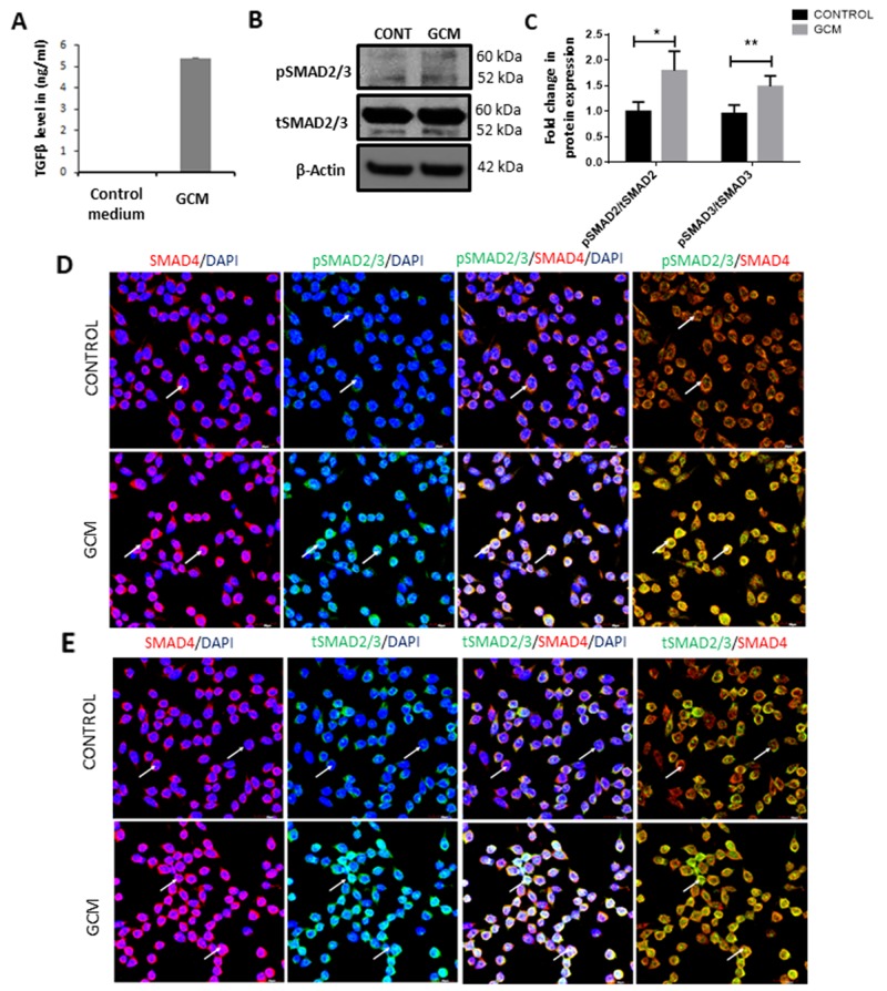
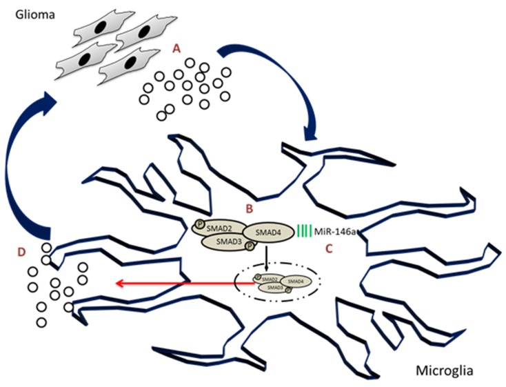
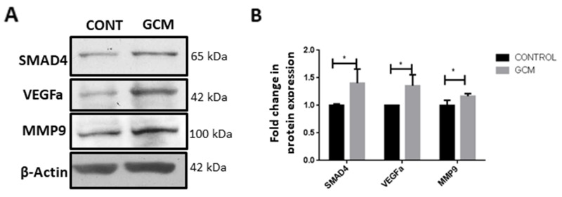
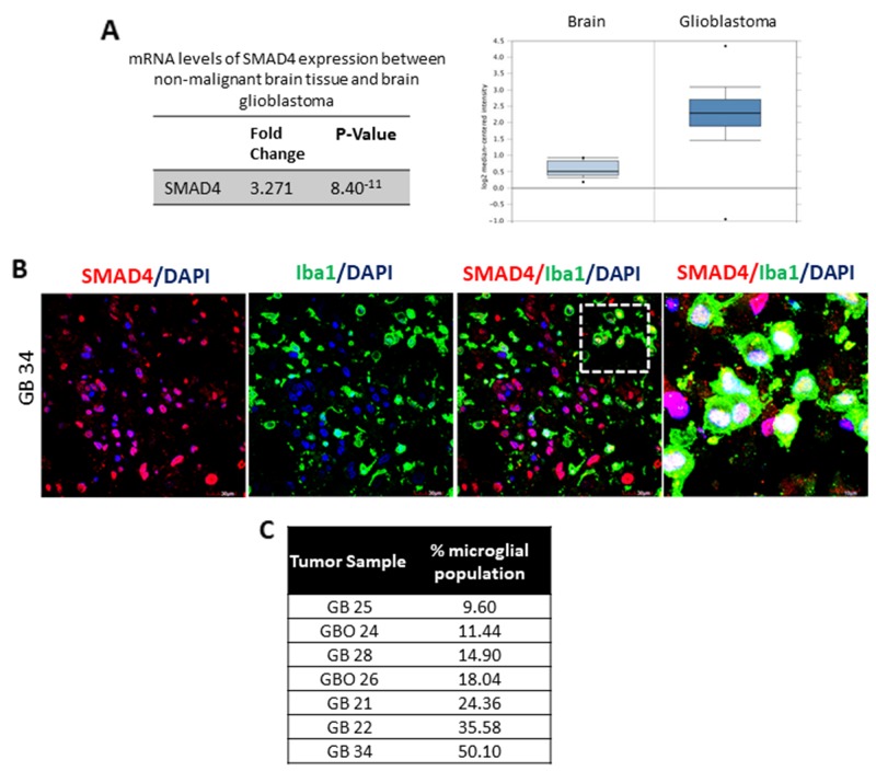
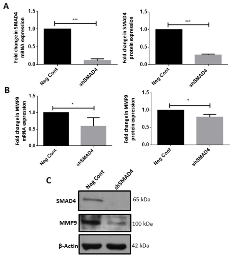
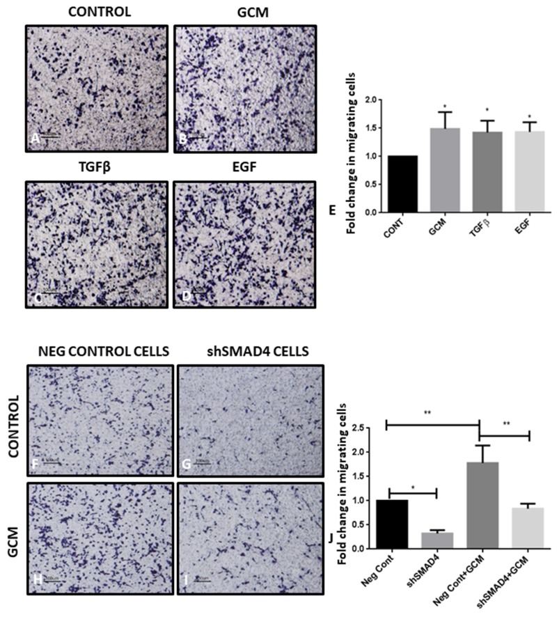
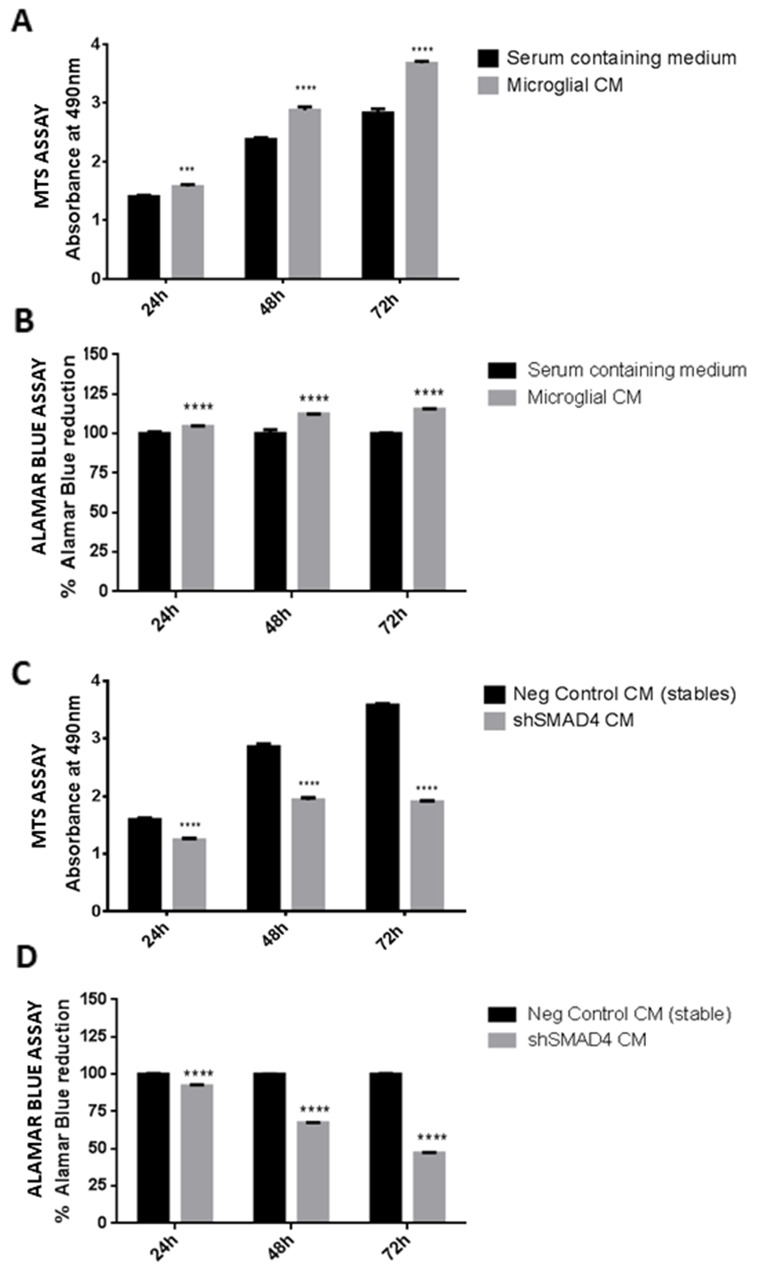
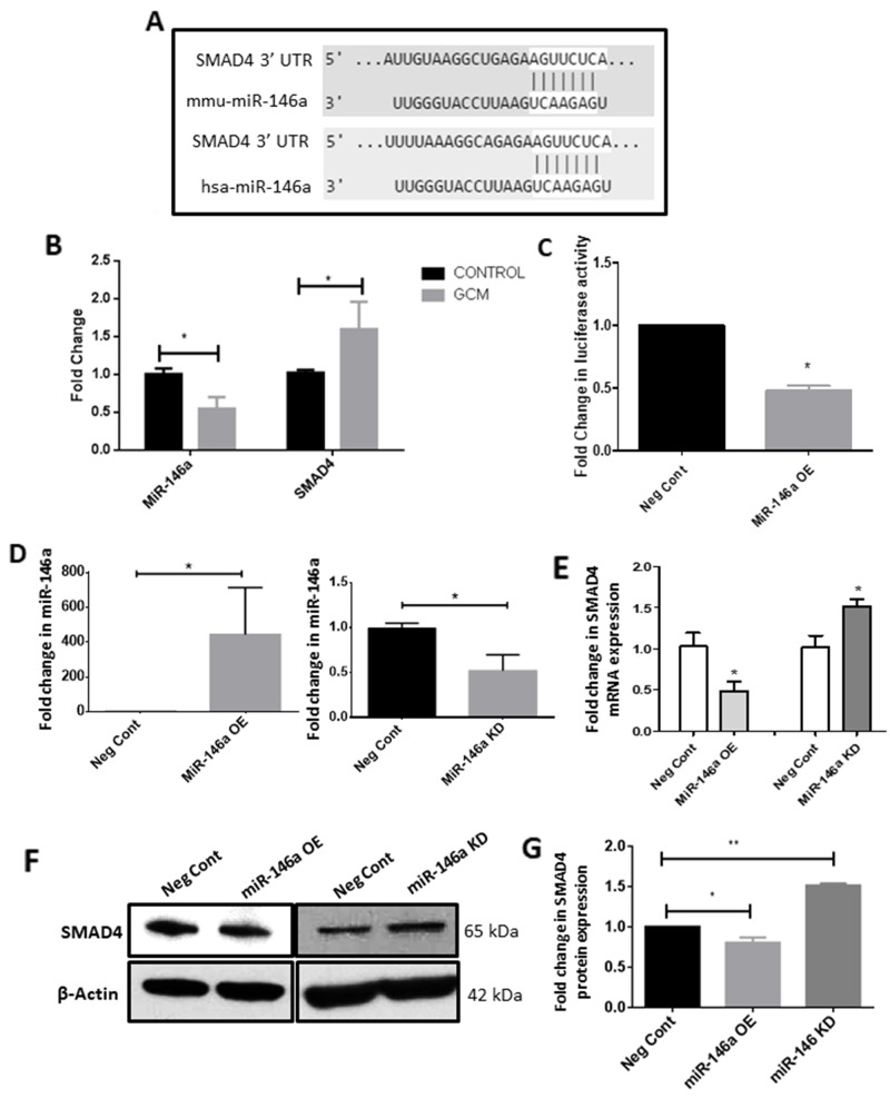
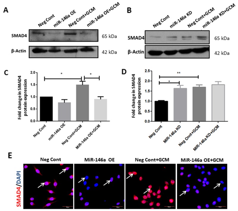
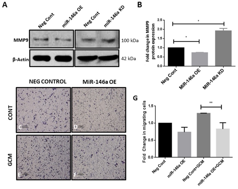
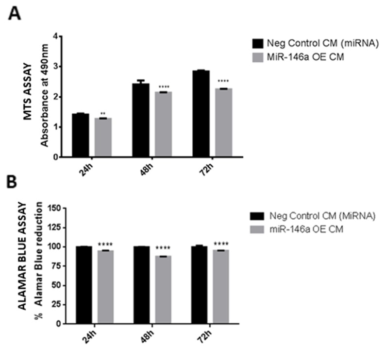
Glioma tumors constitute a significant portion of microglial cells, which are known to support tumor progression. The present study demonstrates that transforming growth factor-β (TGFβ) signaling pathway in microglia in a glioma environment is involved in tumor progression and pathogenesis. It has been shown that the TGFβ level is elevated in higher grades of gliomas and its signaling pathway regulates tumor progression through phosphorylation of SMAD2 and SMAD3, which form a complex with SMAD4 to regulate target gene transcription. In an cell line-based model increased protein levels of pSMAD2/3, total SMAD2/3 and SMAD4 were observed in murine BV2 microglia cultured in glioma conditioned medium (GCM), indicative of the activated TGFβ signaling pathway in microglia associated with glioma environment. Immunofluorescence labeling further revealed the expression of SMAD4 in microglial and non-microglial cells of human glioblastomas tissue . Functional analysis through shRNA-mediated stable knockdown of SMAD4 in microglia revealed the downregulation of the expression of matrix metalloproteinase 9 (MMP9), which has been shown to be involved in tumor progression and cell migration. Further, knockdown of SMAD4 in microglia decreased the migration of microglial cells towards GCM, indicating that SMAD4 promotes microglial migration in glioma environment. In addition, SMAD4 has been shown to be post-transcriptionally regulated by microRNA-146a, which was downregulated in microglia treated with GCM. Overexpression of miR-146a resulted in decreased expression of SMAD4 together with tumor supportive gene MMP9 in microglia, and subsequently suppressed microglial migration towards GCM, possibly through regulation of SMAD4. On the other hand, the cell viability assay revealed decreased viability of glioma cells when they were treated with conditioned medium derived from SMAD4 knockdown microglia or miR-146a overexpressed microglia as compared to glioma cells treated with the medium from control microglial cells. Taken together, the present study suggests that microglial SMAD4 which is epigenetically regulated by miR-146a promotes microglial migration in gliomas and glioma cell viability.
胶质瘤肿瘤构成了小胶质细胞的很大一部分,已知小胶质细胞会促进肿瘤进展。本研究表明,胶质瘤环境中小胶质细胞内的转化生长因子-β(TGFβ)信号通路参与肿瘤进展和发病机制。研究表明,在高级别胶质瘤中TGFβ水平升高,其信号通路通过SMAD2和SMAD3的磷酸化来调节肿瘤进展,SMAD2和SMAD3与SMAD4形成复合物以调节靶基因转录。在基于细胞系的模型中,在胶质瘤条件培养基(GCM)中培养的小鼠BV2小胶质细胞中观察到pSMAD2/3、总SMAD2/3和SMAD4的蛋白水平增加,这表明与胶质瘤环境相关的小胶质细胞中TGFβ信号通路被激活。免疫荧光标记进一步揭示了SMAD4在人胶质母细胞瘤组织的小胶质细胞和非小胶质细胞中的表达。通过shRNA介导的小胶质细胞中SMAD4稳定敲低进行功能分析,结果显示基质金属蛋白酶9(MMP9)的表达下调,MMP9已被证明参与肿瘤进展和细胞迁移。此外,小胶质细胞中SMAD4的敲低减少了小胶质细胞向GCM的迁移,表明SMAD4促进胶质瘤环境中小胶质细胞的迁移。此外,已证明SMAD4受到微小RNA-146a的转录后调控,在用GCM处理的小胶质细胞中微小RNA-146a表达下调。miR-146a的过表达导致小胶质细胞中SMAD4以及肿瘤支持基因MMP9的表达降低,随后抑制了小胶质细胞向GCM的迁移,可能是通过调节SMAD4实现的。另一方面,细胞活力测定显示,与用对照小胶质细胞培养基处理的胶质瘤细胞相比,用来自SMAD4敲低小胶质细胞或miR-146a过表达小胶质细胞的条件培养基处理时,胶质瘤细胞的活力降低。综上所述,本研究表明,受miR-146a表观遗传调控的小胶质细胞SMAD4促进胶质瘤中小胶质细胞的迁移以及胶质瘤细胞的活力。