Thakkar Akanksha N, Chinnadurai Ponraj, Breinholt John P, Lin C Huie
Houston Methodist DeBakey Heart & Vascular Center, Houston Methodist Hospital, Houston, Texas.
Siemens Medical Solutions USA Inc, Hoffman Estates, Illinois.
Catheter Cardiovasc Interv. 2018 Aug 1;92(2):353-357. doi: 10.1002/ccd.27645. Epub 2018 Jun 13.
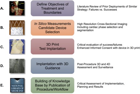
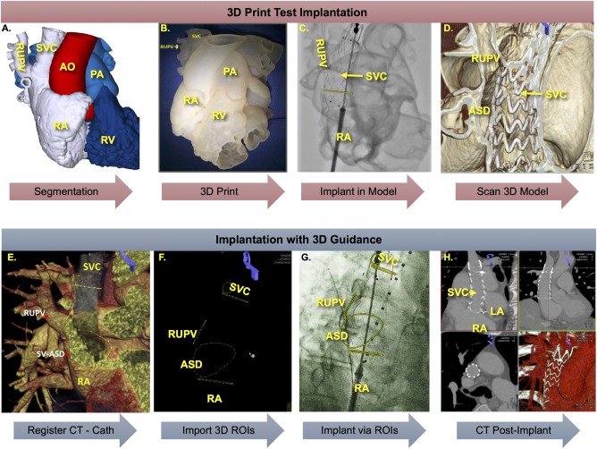
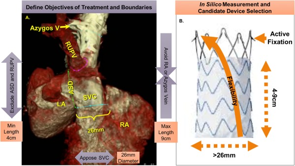
A 63-year-old man with cirrhosis, hepatocellular carcinoma, and coagulopathy was diagnosed with a sinus venosus atrial septal defect (ASD) and partial anomalous pulmonary venous return (PAPVR) of the right upper pulmonary vein (RUPV). Transcatheter repair by positioning a stent graft in the superior vena cava was planned. Based on three-dimensional (3D) reconstruction of gated cardiac CTA, a 28 mm × 7 cm Endurant II aortic extension stent graft (Medtronic, MN) was chosen. A 3D model printed from the CTA was used to simulate device deployment, demonstrating successful exclusion of the sinus venosus ASD with return of the RUPV to the left atrium (LA). Post simulation, the 3D model was used for informed consent. The patient was then taken to the hybrid operating room. On-table cone beam CT was performed and registered with the CTA images. This enabled overlay of 3D regions of interest to live 2D fluoroscopy. The stent graft was then deployed using 3D regions of interest for guidance. Hemodynamics and angiography demonstrated successful exclusion of the sinus venosus ASD and unobstructed return of RUPV to the LA. This is the first report of comprehensive use of contemporary imaging for planning, simulation, patient consent, and procedural guidance for patient-centered complex structural intervention in repair of sinus venosus ASD with PAPVR. We propose this as a process model for continued innovation in structural interventions.
一名患有肝硬化、肝细胞癌和凝血病的63岁男性被诊断出患有静脉窦型房间隔缺损(ASD)和右上肺静脉(RUPV)部分肺静脉异位引流(PAPVR)。计划通过在上腔静脉中放置支架移植物进行经导管修复。基于门控心脏CTA的三维(3D)重建,选择了一个28 mm×7 cm的Endurant II主动脉延长型支架移植物(美敦力公司,明尼苏达州)。从CTA打印出的3D模型用于模拟器械植入,结果显示成功封堵静脉窦型ASD,使RUPV回流至左心房(LA)。模拟后,使用3D模型获取患者知情同意。然后将患者送至杂交手术室。术中进行了锥形束CT检查,并与CTA图像进行配准。这使得能够将3D感兴趣区域叠加到实时二维荧光透视图像上。然后在3D感兴趣区域的引导下植入支架移植物。血流动力学和血管造影显示成功封堵静脉窦型ASD,RUPV顺利回流至LA。这是首次报告在以患者为中心的复杂结构干预修复静脉窦型ASD合并PAPVR中,综合运用现代成像技术进行手术规划、模拟、患者知情同意及手术指导。我们提议将此作为结构干预持续创新的流程模型。