Department of Biomedical Imaging and Image-guided Therapy, Division of Nuclear Medicine, Medical University of Vienna, Waehringer Guertel 18-20, 1090, Vienna, Austria.
Department of Pharmaceutical Technology and Biopharmaceutics, University of Vienna, Vienna, Austria.
Mol Imaging Biol. 2019 Apr;21(2):257-268. doi: 10.1007/s11307-018-1212-0.
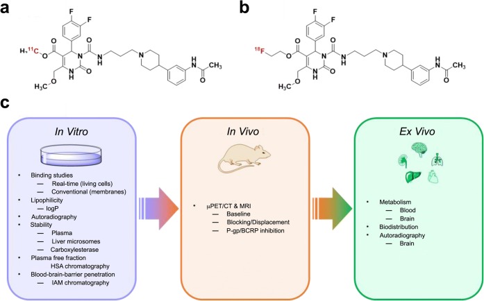
The melanin-concentrating hormone receptor 1 (MCHR1) has become an important pharmacological target, since it may be involved in various diseases, such as diabetes, insulin resistance, and obesity. Hence, a suitable positron emission tomography radiotracer for the in vivo assessment of the MCHR1 pharmacology is imperative. The current paper contrasts the extensive in vitro, in vivo, and ex vivo assessments of the radiotracers [F]FE@SNAP and [C]SNAP-7941 and provides comprehensive information about their biological and physicochemical properties. Furthermore, it examines their suitability for first-in-man imaging studies.
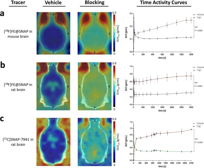
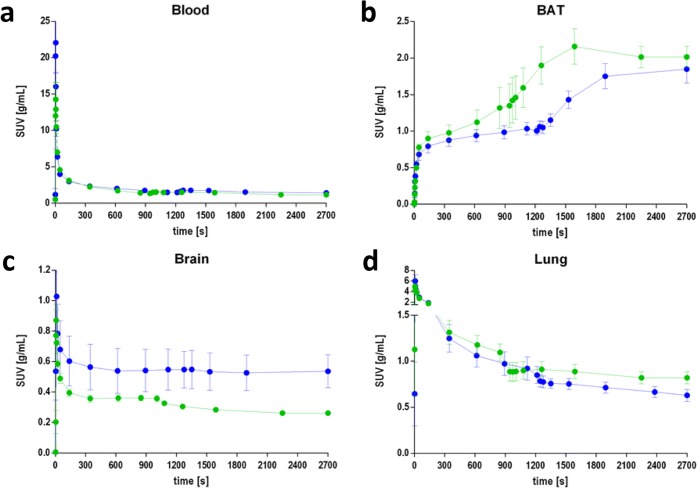
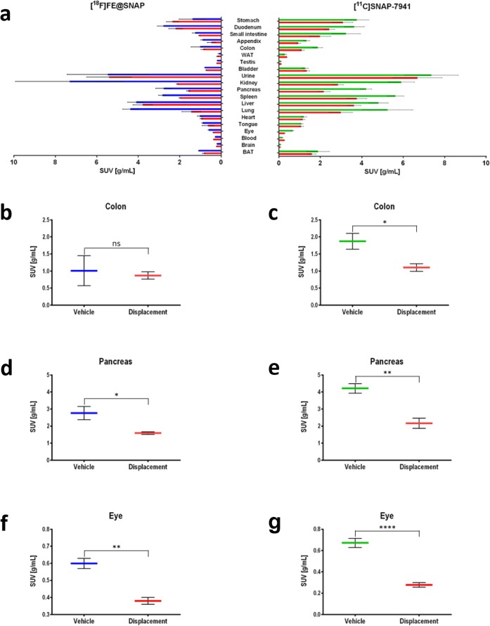
Kinetic real-time cell-binding studies with [F]FE@SNAP and [C]SNAP-7941 were conducted on adherent Chines hamster ovary (CHO-K1) cells stably expressing the human MCHR1 and MCHR2. Small animal imaging studies on mice and rats were performed under displacement and baseline conditions, as well as after pretreatment with the P-glycoprotein/breast cancer resistant protein inhibitor tariquidar. After the imaging studies, detailed analyses of the ex vivo biodistribution were performed. Ex vivo metabolism was determined in rat blood and brain and analyzed at various time points using a quantitative radio-HPLC assay.
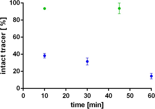
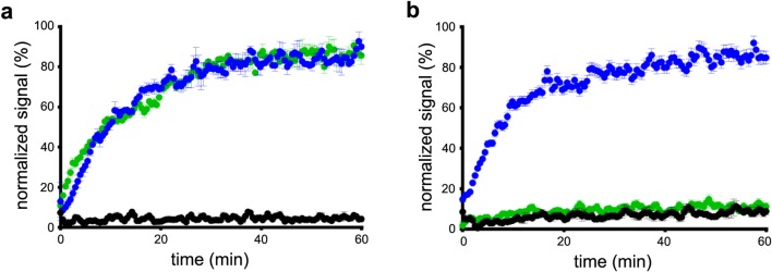
[C]SNAP-7941 demonstrates high uptake on CHO-K1-hMCHR1 cells, whereas no uptake was detected for the CHO-K1-hMCHR2 cells. In contrast, [F]FE@SNAP evinced binding to CHO-K1-hMCHR1 and CHO-K1-hMCHR2 cells. Imaging studies with [F]FE@SNAP and [C]SNAP-7941 showed an increased brain uptake after tariquidar pretreatment in mice, as well as in rats, and exhibited a significant difference between the time-activity curves of the baseline and blocking groups. Biodistribution of both tracers demonstrated a decreased uptake after displacement. [C]SNAP-7941 revealed a high metabolic stability in rats, whereas [F]FE@SNAP was rapidly metabolized.
Both radiotracers demonstrate appropriate imaging properties for the MCHR1. However, the pronounced metabolic stability as well as superior selectivity and affinity of [C]SNAP-7941 underlines the decisive superiority over [F]FE@SNAP.
黑色素浓缩激素受体 1(MCHR1)已成为一个重要的药理学靶点,因为它可能与多种疾病有关,如糖尿病、胰岛素抵抗和肥胖症。因此,需要一种合适的正电子发射断层扫描示踪剂来评估 MCHR1 药理学。本文对比了放射性示踪剂[F]FE@SNAP 和[C]SNAP-7941 的广泛体外、体内和离体评估,并提供了关于其生物学和物理化学特性的综合信息。此外,还考察了它们用于首次人体成像研究的适用性。
采用动力学实时细胞结合研究,在稳定表达人 MCHR1 和 MCHR2 的贴壁中国仓鼠卵巢(CHO-K1)细胞上进行[F]FE@SNAP 和[C]SNAP-7941 的检测。在置换和基础条件下,以及在 P-糖蛋白/乳腺癌耐药蛋白抑制剂 tariquidar 预处理后,在小鼠和大鼠上进行小动物成像研究。在成像研究后,对离体生物分布进行详细分析。在大鼠血液和大脑中进行离体代谢研究,并使用定量放射性 HPLC 分析在不同时间点进行分析。
[C]SNAP-7941 在 CHO-K1-hMCHR1 细胞上表现出高摄取,而在 CHO-K1-hMCHR2 细胞上则没有摄取。相比之下,[F]FE@SNAP 与 CHO-K1-hMCHR1 和 CHO-K1-hMCHR2 细胞结合。[F]FE@SNAP 和[C]SNAP-7941 的成像研究表明,在 tariquidar 预处理后,小鼠和大鼠的大脑摄取增加,并且基础组和阻断组的时间-活性曲线之间存在显著差异。两种示踪剂的生物分布在置换后显示摄取减少。[C]SNAP-7941 在大鼠中表现出较高的代谢稳定性,而[F]FE@SNAP 则迅速代谢。
两种放射性示踪剂均显示出适合 MCHR1 的成像特性。然而,[C]SNAP-7941 的代谢稳定性显著提高,以及在选择性和亲和力方面的优势,凸显了其相对于[F]FE@SNAP 的决定性优势。