Department of Cell, Developmental and Regenerative Biology, Icahn School of Medicine at Mount Sinai, New York, United States.
Elife. 2018 Sep 26;7:e37881. doi: 10.7554/eLife.37881.
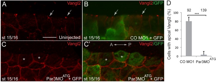
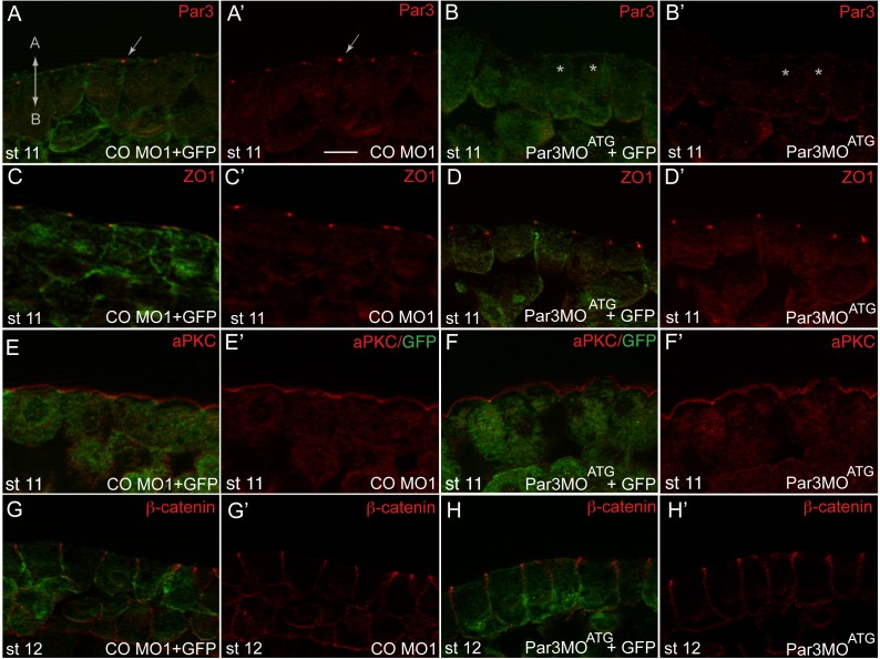
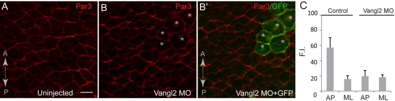
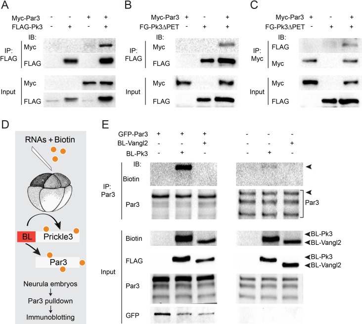
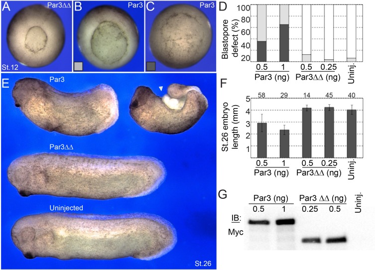
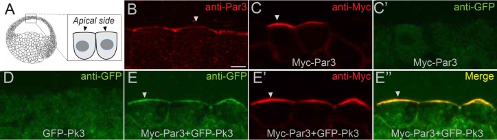
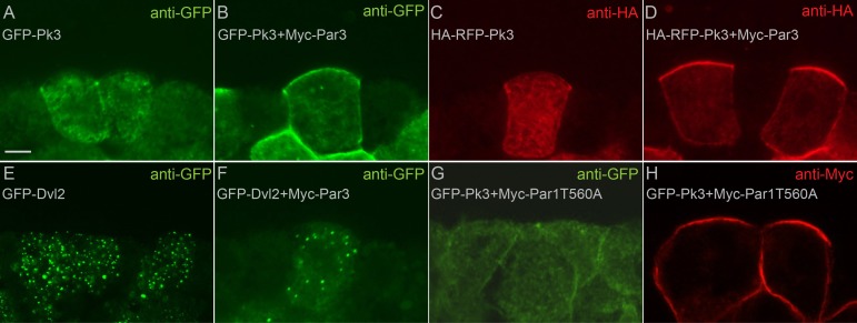
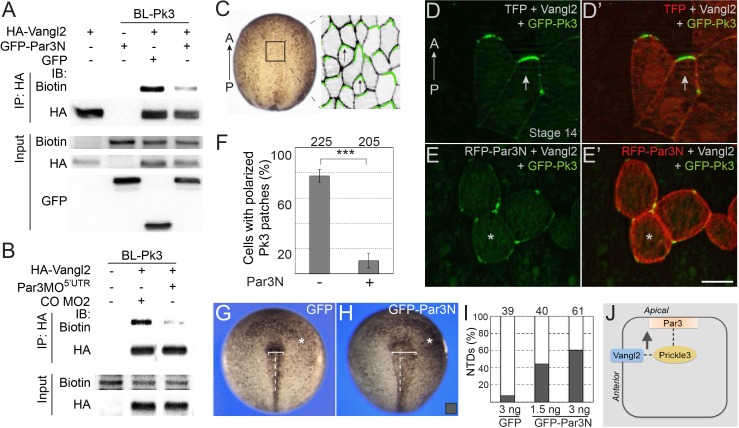
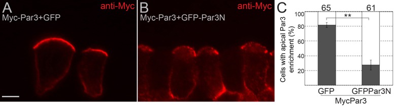
Vertebrate neural tube formation depends on the coordinated orientation of cells in the tissue known as planar cell polarity (PCP). In the neural plate, PCP is marked by the enrichment of the conserved proteins Prickle3 and Vangl2 at anterior cell boundaries. Here we show that the apical determinant Par3 is also planar polarized in the neuroepithelium, suggesting a role for Par3 in PCP. Consistent with this hypothesis, interference with Par3 activity inhibited asymmetric distribution of PCP junctional complexes and caused neural tube defects. Importantly, Par3 physically associated with Prickle3 and promoted its apical localization, whereas overexpression of a Prickle3-binding Par3 fragment disrupted PCP in the neural plate. We also adapted proximity biotinylation assay for use in embryos and show that Par3 functions by enhancing the formation of the anterior apical PCP complex. These findings describe a mechanistic link between the apical localization of PCP components and morphogenetic movements underlying neurulation.
脊椎动物神经管的形成依赖于组织中细胞的协调定向,这个组织被称为平面细胞极性(PCP)。在神经板中,PCP 的特征是保守蛋白 Prickle3 和 Vangl2 在细胞前缘边界的富集。在这里,我们表明顶端决定因素 Par3 在神经上皮中也呈平面极化,这表明 Par3 在 PCP 中起作用。与这一假设一致,干扰 Par3 活性抑制了 PCP 连接复合体的不对称分布,并导致神经管缺陷。重要的是,Par3 与 Prickle3 物理结合,并促进其顶端定位,而 Prickle3 结合的 Par3 片段的过表达破坏了神经板中的 PCP。我们还调整了邻近生物素标记测定法在胚胎中的应用,并表明 Par3 通过增强前顶 PCP 复合体的形成来发挥作用。这些发现描述了 PCP 成分顶端定位与神经管形成的形态发生运动之间的机制联系。