Lee Sanghoon, Zhou Ping, Gupta Anita, Shin Soona
Division of Pediatric General and Thoracic Surgery Cincinnati Children's Hospital Medical Center Cincinnati OH.
Division of Pathology and Laboratory Medicine Cincinnati Children's Hospital Medical Center Cincinnati OH.
Hepatol Commun. 2018 Sep 25;2(10):1199-1212. doi: 10.1002/hep4.1204. eCollection 2018 Oct.
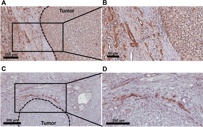
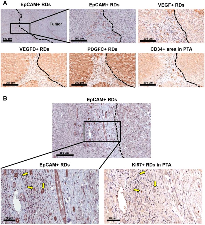
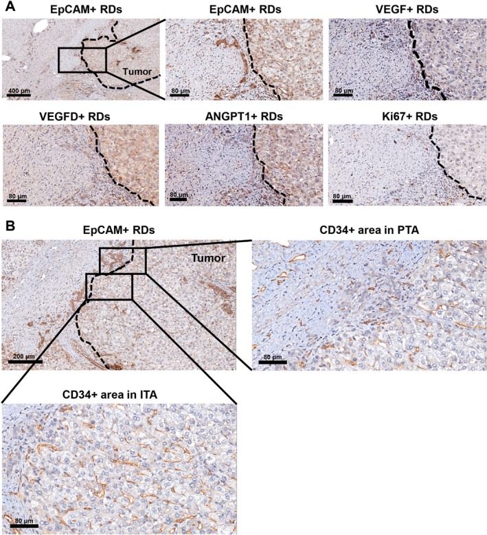
While reactive ductules (RDs) have been observed in viral hepatitis, biliary atresia, nonalcoholic fatty liver disease, and adult hepatocellular carcinoma (HCC), RDs in pediatric liver cancer remain uncharacterized. This study investigated the relationship of RDs with angiogenic paracrine factors, the extent of angiogenesis, and tumor cell proliferation in pediatric hepatoblastoma (HBL)/HCC livers. We quantified the extent of RDs and their expression of paracrine factors that include vascular endothelial growth factor (VEGF), vascular endothelial growth factor D (VEGFD), platelet-derived growth factor C, and angiopoietin 1 (ANGPT1). In addition, we performed immunohistochemical detection of the endothelial marker clusters of differentiation (CD)34 and the proliferation marker Ki67 followed by correlation analyses. In HBL, we found the percentage of RDs with Ki67 expression (% Ki67+ RDs) significantly correlated with intratumoral Ki67+ areas ( 0.5138, 0.0349) and % ANGPT1+ RDs positively correlated with % Ki67+ RDs ( 0.5851, 0.0136). In HCC, the high ANGPT1+ RDs group (i.e., cases with % ANGPT1+ RDs ≥50) exhibited high intratumoral Ki67+ areas compared to the low ANGPT1+ RDs group. In the combined HBL and HCC liver tumor group, there was a positive association between % platelet-derived growth factor C+ RDs and intratumoral Ki67+ areas ( 0.4712, 0.0099) and the high VEGFD+ RDs group (≥50%) exhibited a high number of peritumoral CD34+ vessels compared to the low VEGFD+ RDs group. Paracrine factor-expressing RDs are associated with angiogenesis and proliferation of pediatric liver tumors.
虽然在病毒性肝炎、胆道闭锁、非酒精性脂肪性肝病和成人肝细胞癌(HCC)中已观察到反应性小胆管(RDs),但小儿肝癌中的RDs仍未得到充分描述。本研究调查了小儿肝母细胞瘤(HBL)/HCC肝脏中RDs与血管生成旁分泌因子、血管生成程度和肿瘤细胞增殖之间的关系。我们量化了RDs的程度及其旁分泌因子的表达,这些旁分泌因子包括血管内皮生长因子(VEGF)、血管内皮生长因子D(VEGFD)、血小板衍生生长因子C和血管生成素1(ANGPT1)。此外,我们进行了内皮标志物分化簇(CD)34和增殖标志物Ki67的免疫组化检测,随后进行相关性分析。在HBL中,我们发现Ki67表达的RDs百分比(%Ki67+RDs)与瘤内Ki67+区域显著相关(r = 0.5138,P = 0.0349),%ANGPT1+RDs与%Ki67+RDs呈正相关(r = 0.5851,P = 0.0136)。在HCC中,与低ANGPT1+RDs组相比,高ANGPT1+RDs组(即%ANGPT1+RDs≥50%的病例)表现出较高的瘤内Ki67+区域。在HBL和HCC联合肝脏肿瘤组中,%血小板衍生生长因子C+RDs与瘤内Ki67+区域之间存在正相关(r = 0.4712,P = 0.0099),与低VEGFD+RDs组相比,高VEGFD+RDs组(≥50%)的瘤周CD34+血管数量较多。表达旁分泌因子的RDs与小儿肝脏肿瘤的血管生成和增殖有关。