Bhalchim Supriya Girish, Jugade Shraddha C, Ramaswami Easwaran, Gogri Ajas A, Kadam Sonali G, Umarji Hemant R
Department of Oral Medicine and Radiology, Government Dental College and Hospital, Mumbai, Maharashtra, India.
Contemp Clin Dent. 2018 Sep;9(Suppl 2):S221-S226. doi: 10.4103/ccd.ccd_64_18.
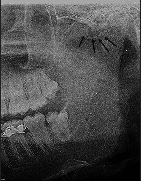
In 1985, Tyndall and Matteson first described the air cells that occur in the root of zygomatic arch and in the articular eminence of the temporal bone but do not extend beyond the zygomaticotemporal suture.
The aim is to study the prevalence and patterns of pneumatized articular tubercle (PAT) retrospectively using two different imaging modalities, i.e., digital panoramic radiography and cone-beam computed tomography (CBCT).
Total 3000 panoramic radiographs belonging to 1291 females and 1709 males and CBCT scans of 200 patients belonging to 62 females and 138 males were studied retrospectively and investigated for radiographic features of pneumatized articular tubercle.
Prevalence found by using panoramic radiography is 1.96% and by using CBCT is 12.5%.
This research scrutinizes the prevalence of pneumatized articular tubercle and establishes the prevalence of the same among the studied population, using panoramic radiography and CBCT.
1985年,廷德尔和马特森首次描述了出现在颧弓根部和颞骨关节结节中但不超过颧颞缝的气房。
旨在使用两种不同的成像方式,即数字化全景X线摄影和锥形束计算机断层扫描(CBCT),回顾性研究气化关节结节(PAT)的患病率和模式。
回顾性研究了属于1291名女性和1709名男性的3000张全景X线片,以及属于62名女性和138名男性的200名患者的CBCT扫描图像,并对气化关节结节的影像学特征进行了研究。
使用全景X线摄影发现的患病率为1.96%,使用CBCT发现的患病率为12.5%。
本研究使用全景X线摄影和CBCT,仔细检查了气化关节结节的患病率,并确定了其在研究人群中的患病率。