• 文献检索
  • 文档翻译
  • 深度研究
  • 学术资讯
  • Suppr Zotero 插件Zotero 插件
  • 邀请有礼
  • 套餐&价格
  • 历史记录
应用&插件
Suppr Zotero 插件Zotero 插件浏览器插件Mac 客户端Windows 客户端微信小程序
定价
高级版会员购买积分包购买API积分包
服务
文献检索文档翻译深度研究API 文档MCP 服务
关于我们
关于 Suppr公司介绍联系我们用户协议隐私条款
关注我们

Suppr 超能文献

核心技术专利:CN118964589B侵权必究
粤ICP备2023148730 号-1Suppr @ 2026

文献检索

告别复杂PubMed语法,用中文像聊天一样搜索,搜遍4000万医学文献。AI智能推荐,让科研检索更轻松。

立即免费搜索

文件翻译

保留排版,准确专业,支持PDF/Word/PPT等文件格式,支持 12+语言互译。

免费翻译文档

深度研究

AI帮你快速写综述,25分钟生成高质量综述,智能提取关键信息,辅助科研写作。

立即免费体验

腺肌瘤样子宫肿瘤的诊断与处理:超声、磁共振成像、手术表现及病理对照

Diagnosis and Management of an Adenomatoid Uterine Tumor: Ultrasound, Magnetic Resonance Imaging, Surgical Appearance, and Pathology Correlation.

作者信息

Orlando Jennie, deRiese Cornelia, Blackwell Eric, Graham Suzanne, Phy Jennifer

机构信息

Department of Obstetrics and Gynecology and Texas Tech University Health Sciences Center, Lubbock, Texas.

Department of Pathology, Texas Tech University Health Sciences Center, Lubbock, Texas.

出版信息

Biores Open Access. 2018 Oct 26;7(1):159-164. doi: 10.1089/biores.2018.0023. eCollection 2018.

DOI:10.1089/biores.2018.0023
PMID:30393587
原文链接:https://pmc.ncbi.nlm.nih.gov/articles/PMC6206546/
Abstract

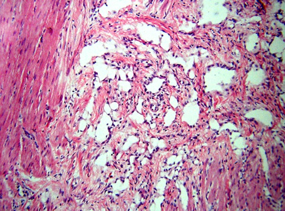
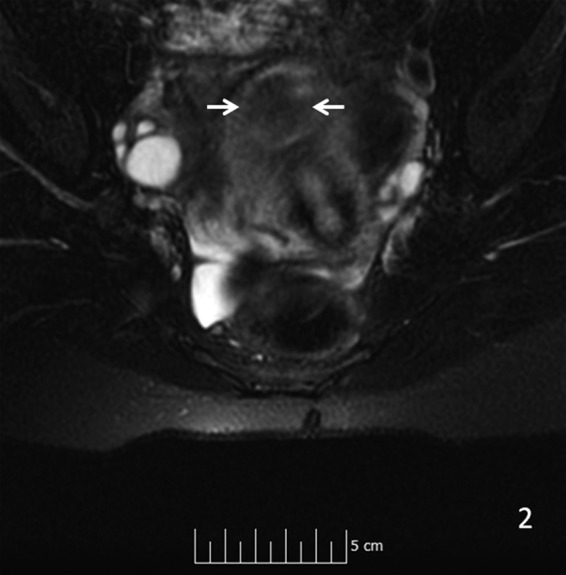
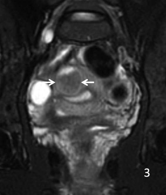
Adenomatoid uterine tumors are rare, and their appearance on medical imaging modalities is not well established. We present a case of an adenomatoid uterine tumor reviewing a unique sonographic presentation, magnetic resonance imaging (MRI), gross surgical appearance of the tumor, and microscopic pathology images. A 29-year-old gravida 0 Caucasian woman presented with dysmenorrhea, menorrhagia, and desire to conceive. Transvaginal ultrasound revealed a 2.7 cm round, well-circumscribed posterior intramural uterine mass. The mass was hyperechoic centrally with a thin hypoechoic rim. Color Doppler imaging revealed a prominent vascular rim around the periphery of the mass as well as central vascularity not typical for a leiomyoma. MRI, with and without intravenous gadolinium, was obtained showing a 2.7 cm posterior fundal mildly enhancing uterine mass suggestive of leiomyoma. The mass had a heterogeneous signal pattern on T2-weighted images, and no fat component was noted within the mass. Repeat transvaginal ultrasound showed interval growth of the mass to 3.5 cm with a lipomatous appearance. Adenomatoid uterine tumors are rare and may be mistaken for uterine leiomyomata. Unique features include sonographic appearance of central hyperechogenicity with a hypoechoic rim and prominent peripheral and central vascularity in conjunction with MRI revealing a heterogeneous signal pattern on T2-weighted images without fat component. Gross surgical appearance reveals a nondiscrete capsule and secretion of mucoid material when the mass is exposed. We present a case of adenomatoid tumor providing sonographic, MRI, surgical, and pathological correlation. The patient subsequently conceived spontaneously and delivered at term by cesarean section. The patient underwent a preoperative evaluation with complete blood count, comprehensive metabolic panel, blood type with antibody screen, and pregnancy test. She underwent laparoscopic excision with robotic assistance for removal of the tumor. Grossly, the uterine mass had a very soft consistency atypical for a uterine leiomyoma making dissection more challenging. During dissection the mass diffusely secreted a mucoid material although the capsule was not disrupted. The lesion was excised intact and was removed from the peritoneal cavity in an endocatch bag without internal morcellation. Microscopic examination revealed an adenomatoid tumor.

摘要

腺肌瘤样子宫肿瘤较为罕见,其在医学影像检查中的表现尚未完全明确。我们报告一例腺肌瘤样子宫肿瘤,回顾其独特的超声表现、磁共振成像(MRI)、肿瘤的大体手术外观及微观病理图像。一名29岁未孕的白人女性,因痛经、月经过多及有生育意愿前来就诊。经阴道超声检查发现一个2.7厘米圆形、边界清晰的子宫肌壁间后壁肿物。肿物中央呈高回声,周边有一薄层低回声边缘。彩色多普勒成像显示肿物周边有明显的血管环,且中央有血管分布,这并非平滑肌瘤的典型表现。行MRI检查,静脉注射钆对比剂前后成像显示一个2.7厘米的子宫底部后壁轻度强化肿物,提示为平滑肌瘤。该肿物在T2加权图像上信号不均匀,且肿物内未发现脂肪成分。再次经阴道超声检查显示肿物增大至3.5厘米,呈脂肪瘤样外观。腺肌瘤样子宫肿瘤较为罕见,可能被误诊为子宫平滑肌瘤。其独特特征包括超声表现为中央高回声、周边低回声边缘及明显的周边和中央血管分布,同时MRI显示T2加权图像上信号不均匀且无脂肪成分。大体手术外观显示肿物无明显包膜,肿物暴露时可见黏液样物质分泌。我们报告一例腺肌瘤样肿瘤,提供超声、MRI、手术及病理的相关性。患者随后自然受孕并足月剖宫产分娩。患者术前进行了全血细胞计数、综合代谢指标检测、血型及抗体筛查和妊娠试验等评估。她在机器人辅助下行腹腔镜肿瘤切除术。大体上,子宫肿物质地非常软,这与子宫平滑肌瘤不同,增加了手术分离的难度。手术分离过程中,肿物弥漫性分泌黏液样物质,尽管包膜未破裂。病变完整切除,装入内套袋从腹腔取出,未进行内部碎解。显微镜检查显示为腺肌瘤样肿瘤。

https://cdn.ncbi.nlm.nih.gov/pmc/blobs/6de7/6206546/1b846ff57898/fig-10.jpg
https://cdn.ncbi.nlm.nih.gov/pmc/blobs/6de7/6206546/8e74382e252d/fig-1.jpg
https://cdn.ncbi.nlm.nih.gov/pmc/blobs/6de7/6206546/92f5e11994c8/fig-2.jpg
https://cdn.ncbi.nlm.nih.gov/pmc/blobs/6de7/6206546/0878073ffb1f/fig-3.jpg
https://cdn.ncbi.nlm.nih.gov/pmc/blobs/6de7/6206546/261162d2be97/fig-4.jpg
https://cdn.ncbi.nlm.nih.gov/pmc/blobs/6de7/6206546/06b8a73e083d/fig-5.jpg
https://cdn.ncbi.nlm.nih.gov/pmc/blobs/6de7/6206546/659c00bc98b3/fig-6.jpg
https://cdn.ncbi.nlm.nih.gov/pmc/blobs/6de7/6206546/55efe4613dbd/fig-7.jpg
https://cdn.ncbi.nlm.nih.gov/pmc/blobs/6de7/6206546/b81bf879ffac/fig-8.jpg
https://cdn.ncbi.nlm.nih.gov/pmc/blobs/6de7/6206546/bbdcd9b2ea27/fig-9.jpg
https://cdn.ncbi.nlm.nih.gov/pmc/blobs/6de7/6206546/1b846ff57898/fig-10.jpg

相似文献

1
Diagnosis and Management of an Adenomatoid Uterine Tumor: Ultrasound, Magnetic Resonance Imaging, Surgical Appearance, and Pathology Correlation.腺肌瘤样子宫肿瘤的诊断与处理:超声、磁共振成像、手术表现及病理对照
Biores Open Access. 2018 Oct 26;7(1):159-164. doi: 10.1089/biores.2018.0023. eCollection 2018.
2
A subserosal, pedunculated, multilocular uterine leiomyoma with ovarian tumor-like morphology and histological architecture of adenomatoid tumors: a case report and review of the literature.一例具有腺瘤样瘤的卵巢肿瘤样形态和组织学结构的浆膜下带蒂多房性子宫平滑肌瘤:病例报告及文献复习
J Med Case Rep. 2016 Dec 20;10(1):352. doi: 10.1186/s13256-016-1167-1.
3
Uterine-sparing Laparoscopic Resection of Accessory Cavitated Uterine Masses.保留子宫的腹腔镜切除附件囊性子宫肿块。
J Minim Invasive Gynecol. 2018 Jan;25(1):24-25. doi: 10.1016/j.jmig.2017.06.001. Epub 2017 Jul 21.
4
MR Imaging Findings of Uterine Adenomatoid Tumors.子宫腺瘤样肿瘤的磁共振成像表现。
Magn Reson Med Sci. 2024 Apr 1;23(2):127-135. doi: 10.2463/mrms.mp.2022-0067. Epub 2023 Jan 26.
5
The management of uterine fibroids in women with otherwise unexplained infertility.患有不明原因不孕症的女性子宫肌瘤的管理。
J Obstet Gynaecol Can. 2015 Mar;37(3):277-285. doi: 10.1016/S1701-2163(15)30318-2.
6
Evaluation of Red Degeneration of Uterine Leiomyoma with Susceptibility-weighted MR Imaging.磁共振磁敏感加权成像评价子宫肌瘤红色变性。
Magn Reson Med Sci. 2019 Apr 10;18(2):158-162. doi: 10.2463/mrms.mp.2018-0074. Epub 2018 Oct 1.
7
Spontaneous uterine rupture in the 33rd week of IVF pregnancy after laparoscopically assisted enucleation of uterine adenomatoid tumor.腹腔镜辅助下子宫腺瘤样瘤摘除术后体外受精妊娠第33周时发生自发性子宫破裂。
J Obstet Gynaecol Res. 2011 May;37(5):452-7. doi: 10.1111/j.1447-0756.2010.01361.x. Epub 2011 Jan 5.
8
Case 225: Leiomyoma.病例 225:平滑肌瘤。
Radiology. 2016 Jan;278(1):297-301. doi: 10.1148/radiol.2015130251.
9
Multicentric infarcted leiomyoadenomatoid tumor: a case report.多中心梗死性平滑肌瘤样肿瘤:一例报告
Int J Clin Exp Pathol. 2009;2(1):99-103. Epub 2008 Feb 11.
10
Management of a giant uterine smooth muscle tumor of uncertain malignant potential in a 32-year-old woman: case report and review of the literature.一名32岁女性巨大子宫平滑肌肿瘤(恶性潜能不确定)的管理:病例报告及文献综述
Minerva Obstet Gynecol. 2022 Oct;74(5):466-470. doi: 10.23736/S2724-606X.22.05126-0. Epub 2022 Aug 1.

引用本文的文献

1
Adenomatoid tumors of ovary mimicking malignancy: report of 2 cases and literature review.卵巢腺瘤样瘤酷似恶性肿瘤:2 例报告及文献复习。
BMC Womens Health. 2022 Dec 26;22(1):547. doi: 10.1186/s12905-022-02138-6.

本文引用的文献

1
True incidence of uterine adenomatoid tumors.子宫腺瘤样瘤的真实发病率。
Biomed Rep. 2013 May;1(3):352-354. doi: 10.3892/br.2013.72. Epub 2013 Mar 5.
2
Adenomatoid Tumors of the Genital Tract.生殖道腺瘤样瘤
Am J Pathol. 1945 Jan;21(1):63-79.
3
Immunoperoxidase localization of keratin proteins, carcinoembryonic antigen, and factor VIII in adenomatoid tumors: evidence for a mesothelial derivation.
Hum Pathol. 1982 Dec;13(12):1106-8. doi: 10.1016/s0046-8177(82)80247-5.