Abdullah Sharif, Guo Ming Hao, Darling Gail, Patsios Demetris
Joint Department of Medical Imaging, University Health Network, Toronto, Canada.
Faculty of Medicine, University Toronto, Toronto, Canada.
BJR Case Rep. 2016 Nov 2;2(4):20150469. doi: 10.1259/bjrcr.20150469. eCollection 2016.
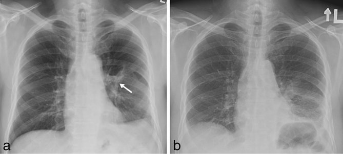
We present a unique case of intercostal muscle flap (ICMF) ossification mimicking an intrathoracic rib diagnosed 3 years after oesophageal perforation repair. A 58-year-old male presented with complaints of mild chest discomfort. Three years ago he had undergone left thoracotomy and primary repair of post-emetic oesophageal perforation. An ICMF had been used to strengthen the repair. Chest X-ray identified a linear calcific density within the left hemithorax. Subsequent thoracic CT characterized the anomaly as ossification of the ICMF. The lesion had the appearance of a well-differentiated intrathoracic rib coursing through the left lower lobe. We discuss the typical appearances of ossified ICMFs and the potential complications resulting from this ossification.
我们报告了一例独特的肋间肌瓣(ICMF)骨化病例,该病例在食管穿孔修复3年后被诊断为酷似胸内肋骨。一名58岁男性因轻度胸部不适前来就诊。三年前,他接受了左胸切开术及催吐后食管穿孔的一期修复。曾使用ICMF加强修复。胸部X线检查发现左半胸内有一线状钙化密度影。随后的胸部CT检查将该异常特征为ICMF骨化。该病变表现为一条分化良好的胸内肋骨贯穿左下叶。我们讨论了骨化ICMF的典型表现以及这种骨化可能导致的并发症。