• 文献检索
  • 文档翻译
  • 深度研究
  • 学术资讯
  • Suppr Zotero 插件Zotero 插件
  • 邀请有礼
  • 套餐&价格
  • 历史记录
应用&插件
Suppr Zotero 插件Zotero 插件浏览器插件Mac 客户端Windows 客户端微信小程序
定价
高级版会员购买积分包购买API积分包
服务
文献检索文档翻译深度研究API 文档MCP 服务
关于我们
关于 Suppr公司介绍联系我们用户协议隐私条款
关注我们

Suppr 超能文献

核心技术专利:CN118964589B侵权必究
粤ICP备2023148730 号-1Suppr @ 2026

文献检索

告别复杂PubMed语法,用中文像聊天一样搜索,搜遍4000万医学文献。AI智能推荐,让科研检索更轻松。

立即免费搜索

文件翻译

保留排版,准确专业,支持PDF/Word/PPT等文件格式,支持 12+语言互译。

免费翻译文档

深度研究

AI帮你快速写综述,25分钟生成高质量综述,智能提取关键信息,辅助科研写作。

立即免费体验

四维计算机断层扫描得出的左心耳射血分数对预测瓣膜性心脏病患者血栓栓塞风险的益处。

Benefit of Four-Dimensional Computed Tomography Derived Ejection Fraction of the Left Atrial Appendage to Predict Thromboembolic Risk in the Patients with Valvular Heart Disease.

作者信息

Kim In Cheol, Chang Hyuk Jae, Cho In Jeong, Shim Chi Young, Hong Geu Ru, Heo Ji Hoe, Nam Hyo Suk, Kim Young Jin, Choi Byoung Wook, Chung Namsik

机构信息

Division of Cardiology, Severance Cardiovascular Hospital, Yonsei University College of Medicine, Seoul, Korea.

Division of Cardiology, Department of Internal Medicine, Dongsan Medical Center, Keimyung University College of Medicine, Daegu, Korea.

出版信息

Korean Circ J. 2019 Feb;49(2):173-180. doi: 10.4070/kcj.2018.0152. Epub 2018 Nov 5.

DOI:10.4070/kcj.2018.0152
PMID:30468031
原文链接:https://pmc.ncbi.nlm.nih.gov/articles/PMC6351282/
Abstract

BACKGROUND AND OBJECTIVES

Decreased left atrial appendage (LAA) emptying velocity in transesophageal echocardiography (TEE) is related with higher incidence of thrombus and increased risk of stroke. Patients with valve disease are at higher risk of thrombus formation before and after surgery. The aim of this study was to investigate the role of 4-dimensional cardiac computed tomography (4DCT) to predict the risk of thrombus formation.

METHODS

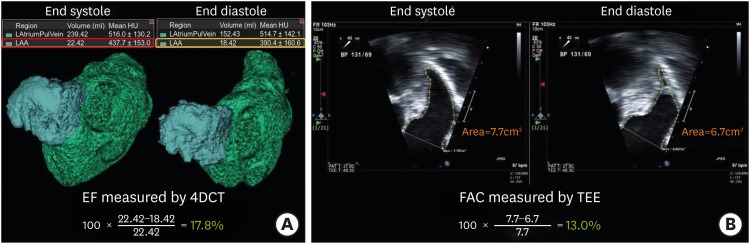
Between March 2010 to March 2015, total of 62 patients (mean 60±15 years old, male: 53.2%) who underwent 4DCT and TEE for cardiac valve evaluation before surgery were retrospectively included in the current study. Fractional area change in TEE view and emptying velocity at left atrial appendage in TEE view (Ve) were measured. Ejection fraction (EF) of left atrial appendage in computed tomography (EF) was calculated by 4DCT with full volume analysis. The best cut-off value of EF predicting presence of spontaneous echo contrast (SEC) or thrombus was evaluated, and correlation between the parameters were also estimated.

RESULTS

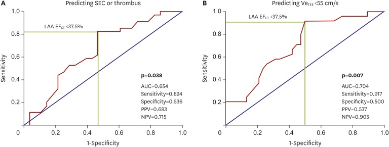
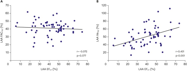
SEC or thrombus was observed in 45.2%. EF and Ve were significantly correlated (r=0.452, p<0.001). However, fractional area change measured by TEE showed no correlation with Ve (r=0.085, p=0.512). EF <37.5% best predicted SEC or thrombus in the patients with valve disease who underwent 4DCT and TEE (area under the curve, 0.654; p=0.038).

CONCLUSIONS

In the patients who underwent 4DCT for cardiac valve evaluation before surgery, EF by volume analysis might have additional role to evaluate LAA function and estimate the risk of thrombus.

摘要

背景与目的

经食管超声心动图(TEE)检查发现左心耳(LAA)排空速度降低与血栓形成发生率较高及中风风险增加相关。瓣膜病患者在手术前后发生血栓形成的风险较高。本研究旨在探讨四维心脏计算机断层扫描(4DCT)在预测血栓形成风险中的作用。

方法

回顾性纳入2010年3月至2015年3月期间62例(平均年龄60±15岁,男性占53.2%)在手术前行4DCT和TEE进行心脏瓣膜评估的患者。测量TEE视图中的面积变化分数以及TEE视图中左心耳的排空速度(Ve)。通过4DCT全容积分析计算计算机断层扫描中左心耳的射血分数(EF)。评估预测自发回声增强(SEC)或血栓存在的EF最佳截断值,并估计各参数之间的相关性。

结果

观察到45.2%的患者存在SEC或血栓。EF与Ve显著相关(r = 0.452,p < 0.001)。然而,TEE测量的面积变化分数与Ve无相关性(r = 0.085,p = 0.512)。EF < 37.5%对接受4DCT和TEE检查的瓣膜病患者的SEC或血栓具有最佳预测价值(曲线下面积,0.654;p = 0.038)。

结论

对于手术前行4DCT进行心脏瓣膜评估的患者,通过容积分析得出的EF可能在评估LAA功能和估计血栓风险方面具有额外作用。

https://cdn.ncbi.nlm.nih.gov/pmc/blobs/b260/6351282/79aa1795068b/kcj-49-173-g003.jpg
https://cdn.ncbi.nlm.nih.gov/pmc/blobs/b260/6351282/01106fe1b542/kcj-49-173-g001.jpg
https://cdn.ncbi.nlm.nih.gov/pmc/blobs/b260/6351282/801ea3262575/kcj-49-173-g002.jpg
https://cdn.ncbi.nlm.nih.gov/pmc/blobs/b260/6351282/79aa1795068b/kcj-49-173-g003.jpg
https://cdn.ncbi.nlm.nih.gov/pmc/blobs/b260/6351282/01106fe1b542/kcj-49-173-g001.jpg
https://cdn.ncbi.nlm.nih.gov/pmc/blobs/b260/6351282/801ea3262575/kcj-49-173-g002.jpg
https://cdn.ncbi.nlm.nih.gov/pmc/blobs/b260/6351282/79aa1795068b/kcj-49-173-g003.jpg

相似文献

1
Benefit of Four-Dimensional Computed Tomography Derived Ejection Fraction of the Left Atrial Appendage to Predict Thromboembolic Risk in the Patients with Valvular Heart Disease.四维计算机断层扫描得出的左心耳射血分数对预测瓣膜性心脏病患者血栓栓塞风险的益处。
Korean Circ J. 2019 Feb;49(2):173-180. doi: 10.4070/kcj.2018.0152. Epub 2018 Nov 5.
2
Predictive value of left atrial appendage lobes on left atrial thrombus or spontaneous echo contrast in patients with non-valvular atrial fibrillation.非瓣膜性心房颤动患者左心耳叶对左心房血栓或自发显影的预测价值
BMC Cardiovasc Disord. 2018 Jul 31;18(1):153. doi: 10.1186/s12872-018-0889-y.
3
Multidetector row computed tomography for identification of left atrial appendage filling defects in patients undergoing pulmonary vein isolation for treatment of atrial fibrillation: comparison with transesophageal echocardiography.多排螺旋计算机断层扫描用于识别接受肺静脉隔离治疗心房颤动患者的左心耳充盈缺损:与经食管超声心动图的比较
Heart Rhythm. 2008 Feb;5(2):253-60. doi: 10.1016/j.hrthm.2007.10.025. Epub 2007 Oct 18.
4
Assessment of left atrial appendage function by transthoracic pulsed Doppler echocardiography: Comparing against transesophageal interrogation and predicting echocardiographic risk factors for stroke.经胸脉冲多普勒超声心动图评估左心耳功能:与经食管检查对比并预测中风的超声心动图危险因素
Echocardiography. 2017 Oct;34(10):1478-1485. doi: 10.1111/echo.13659. Epub 2017 Aug 28.
5
Left atrial appendage filling defects identified by multidetector computed tomography in patients undergoing radiofrequency pulmonary vein antral isolation: a comparison with transesophageal echocardiography.多排螺旋计算机断层扫描在接受射频肺静脉前庭隔离术患者中发现的左心耳充盈缺损:与经食管超声心动图的比较
Am Heart J. 2007 Dec;154(6):1199-205. doi: 10.1016/j.ahj.2007.08.004. Epub 2007 Sep 14.
6
[Stratification of the thromboembolic risk in patients with non-rheumatic atrial fibrillation: assessment of left atrial dysfunction].[非风湿性心房颤动患者血栓栓塞风险的分层:左心房功能障碍的评估]
G Ital Cardiol. 1996 Mar;26(3):273-85.
7
Evaluation of left atrial appendage functions in patients with thrombus and spontaneous echo contrast in left atrial appendage by using color Doppler tissue imaging.应用彩色多普勒组织成像评估左心耳有血栓及自发显影的患者左心耳功能。
Ann Noninvasive Electrocardiol. 2004 Oct;9(4):345-51. doi: 10.1111/j.1542-474X.2004.94573.x.
8
Left atrial appendage 2D-strain assessed by transesophageal echocardiography is associated with thromboembolic risk in patients with atrial fibrillation.经食管超声心动图评估的左心耳二维应变与心房颤动患者的血栓栓塞风险相关。
Turk Kardiyol Dern Ars. 2019 Mar;47(2):111-121. doi: 10.5543/tkda.2019.39482.
9
Cardiac CT Differentiates Left Atrial Appendage Thrombi from Circulatory Stasis in Acute Stroke Patients.心脏CT鉴别急性卒中患者左心耳血栓与循环淤滞
In Vivo. 2016;30(5):671-6.
10
Left atrial appendage emptying fraction assessed by a feature-tracking echocardiographic method is a determinant of thrombus in patients with nonvalvular atrial fibrillation.经特征追踪超声心动图方法评估的左心耳排空分数是无瓣膜性心房颤动患者血栓形成的决定因素。
J Cardiol. 2012 May;59(3):329-36. doi: 10.1016/j.jjcc.2012.01.002. Epub 2012 Feb 17.

引用本文的文献

1
Atrial fibrillation and thromboembolic risk in hypertrophic cardiomyopathy.肥厚型心肌病中的心房颤动与血栓栓塞风险
J Cardiovasc Imaging. 2025 Aug 25;33(1):12. doi: 10.1186/s44348-025-00057-2.
2
Morphological and functional assessment of the left atrial appendage in daily practice: a comprehensive approach using basic and advanced echocardiography with practical tips.日常实践中左心耳的形态学和功能评估:一种使用基础和高级超声心动图的综合方法及实用技巧
J Cardiovasc Imaging. 2024 Jul 29;32(1):12. doi: 10.1186/s44348-024-00017-2.
3
[Computed Tomography of the Left Atrium and Left Atrial Appendage: A Pictorial Essay on the Anatomy, Normal Variants, and Pathology].

本文引用的文献

1
Comparison of Cardiac Computed Tomography With Transesophageal Echocardiography for Identifying Vegetation and Intracardiac Complications in Patients With Infective Endocarditis in the Era of 3-Dimensional Images.比较三维成像时代经胸超声心动图与心脏计算机断层扫描在感染性心内膜炎患者中识别赘生物和心内并发症的价值。
Circ Cardiovasc Imaging. 2018 Mar;11(3):e006986. doi: 10.1161/CIRCIMAGING.117.006986.
2
Left Atrial Appendage Thrombus in Transcatheter Aortic Valve Replacement: Incidence, Clinical Impact, and the Role of Cardiac Computed Tomography.经导管主动脉瓣置换术后左心耳血栓:发生率、临床影响和心脏计算机断层扫描的作用。
JACC Cardiovasc Interv. 2017 Jan 23;10(2):176-184. doi: 10.1016/j.jcin.2016.10.043.
3
[左心房及左心耳的计算机断层扫描:关于解剖结构、正常变异及病理学的图文论述]
Taehan Yongsang Uihakhoe Chi. 2020 Mar;81(2):272-289. doi: 10.3348/jksr.2020.81.2.272. Epub 2020 Mar 31.
4
Multidimensional Approach of Heart Failure Diagnosis and Prognostication Utilizing Cardiac Imaging with Biomarkers.利用心脏成像与生物标志物的心力衰竭诊断和预后评估的多维方法。
Diagnostics (Basel). 2022 Jun 1;12(6):1366. doi: 10.3390/diagnostics12061366.
5
Simultaneous Functional and Morphological Assessment of Left Atrial Appendage by 3D Virtual Models.利用 3D 虚拟模型对左心耳进行功能和形态的同步评估。
J Healthc Eng. 2019 May 21;2019:7095845. doi: 10.1155/2019/7095845. eCollection 2019.
6
Computed Tomography for Assessment of Left Atrial Appendage Function.用于评估左心耳功能的计算机断层扫描
Korean Circ J. 2019 Feb;49(2):181-182. doi: 10.4070/kcj.2018.0355. Epub 2018 Nov 7.
Intensity of Left Atrial Spontaneous Echo Contrast as a Correlate for Stroke Risk Stratification in Patients with Nonvalvular Atrial Fibrillation.
非瓣膜性心房颤动患者左心房自发显影强度与卒中风险分层的相关性
Sci Rep. 2016 Jun 9;6:27650. doi: 10.1038/srep27650.
4
Poor Outcome of Stroke Patients With Atrial Fibrillation in the Presence of Coexisting Spontaneous Echo Contrast.存在自发显影对比的心房颤动中风患者预后不良。
Stroke. 2016 Jul;47(7):1920-2. doi: 10.1161/STROKEAHA.116.013351. Epub 2016 May 17.
5
Potential Utility of Multidetector Computed Tomography to Identify both Cardiac Embolic Sources and Coronary Artery Disease in Patients with Embolic Stroke.多排螺旋计算机断层扫描在识别栓塞性卒中患者心脏栓子来源和冠状动脉疾病方面的潜在效用
Cardiology. 2016;133(4):205-10. doi: 10.1159/000441277. Epub 2015 Dec 1.
6
Stroke after aortic valve surgery: results from a prospective cohort.主动脉瓣置换术后的卒中:一项前瞻性队列研究结果
Circulation. 2014 Jun 3;129(22):2253-61. doi: 10.1161/CIRCULATIONAHA.113.005084. Epub 2014 Apr 1.
7
Prognostic significance of spontaneous echocardiographic contrast detected by transthoracic and transesophageal echocardiography in the era of harmonic imaging.经胸超声心动图和经食管超声心动图检测自发性超声对比在谐波成像时代的预后意义。
Arch Med Sci. 2013 Oct 31;9(5):808-14. doi: 10.5114/aoms.2013.38674. Epub 2013 Nov 5.
8
Guidelines for performing a comprehensive transesophageal echocardiographic examination: recommendations from the American Society of Echocardiography and the Society of Cardiovascular Anesthesiologists.经食管超声心动图全面检查操作指南:美国超声心动图学会和心血管麻醉医师学会的建议
J Am Soc Echocardiogr. 2013 Sep;26(9):921-64. doi: 10.1016/j.echo.2013.07.009.
9
Detection of left atrial appendage thrombus by cardiac computed tomography in patients with atrial fibrillation: a meta-analysis.经心脏计算机断层扫描检测心房颤动患者左心耳血栓:一项荟萃分析。
Circ Cardiovasc Imaging. 2013 Mar 1;6(2):185-94. doi: 10.1161/CIRCIMAGING.112.000153. Epub 2013 Feb 13.
10
Cross-sectional computed tomographic assessment improves accuracy of aortic annular sizing for transcatheter aortic valve replacement and reduces the incidence of paravalvular aortic regurgitation.经胸超声心动图评估改善了经导管主动脉瓣置换术主动脉瓣环尺寸测量的准确性,并降低了瓣周主动脉瓣反流的发生率。
J Am Coll Cardiol. 2012 Apr 3;59(14):1275-86. doi: 10.1016/j.jacc.2011.11.045. Epub 2012 Feb 22.