Kurowski Agata, Molotkov Andrei, Soriano Philippe
Department of Cell, Developmental, and Regenerative Biology, Icahn School of Medicine at Mount Sinai, New York, NY 10029, United States.
Department of Cell, Developmental, and Regenerative Biology, Icahn School of Medicine at Mount Sinai, New York, NY 10029, United States.
Dev Biol. 2019 Feb 1;446(1):94-101. doi: 10.1016/j.ydbio.2018.12.008. Epub 2018 Dec 12.
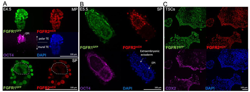
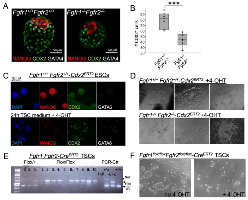
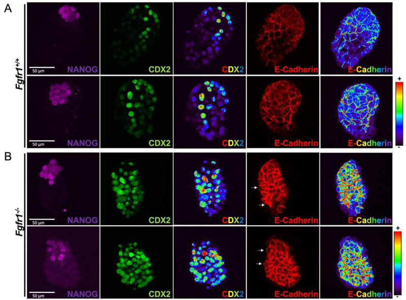
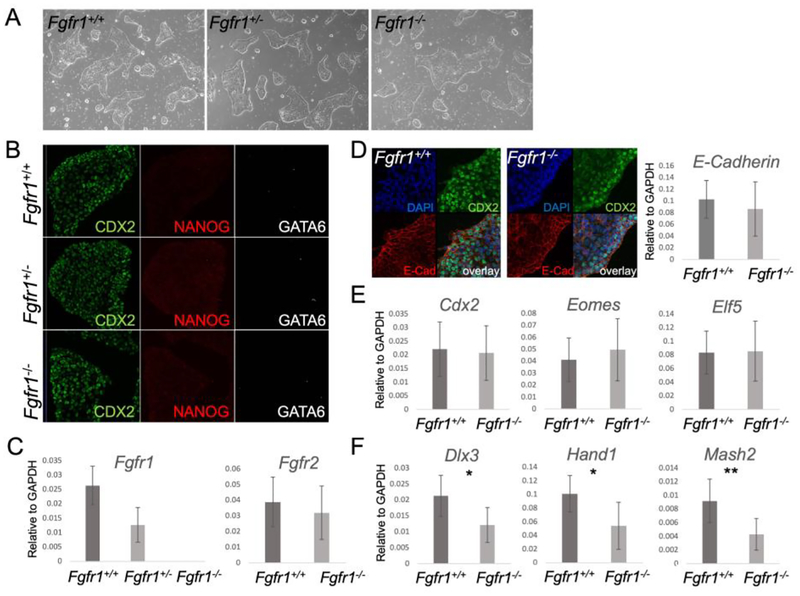
FGF signaling plays important roles in many aspects of mammalian development. Fgfr1 and Fgfr1Fgfr2 mouse embryos on a 129S4 co-isogenic background fail to survive past the peri-implantation stage, whereas Fgfr2 embryos die at midgestation and show defects in limb and placental development. To investigate the basis for the Fgfr1 and Fgfr1Fgfr2 peri-implantation lethality, we examined the role of FGFR1 and FGFR2 in trophectoderm (TE) development. In vivo, Fgfr1 TE cells failed to downregulate CDX2 in the mural compartment and exhibited abnormal apicobasal E-Cadherin polarity. In vitro, we were able to derive mutant trophoblast stem cells (TSCs) from Fgfr1 or Fgfr2 single mutant, but not from Fgfr1Fgfr2 double mutant blastocysts. Fgfr1 TSCs however failed to efficiently upregulate TE differentiation markers upon differentiation. These results suggest that while the TE is specified in Fgfr1 mutants, its differentiation abilities are compromised leading to defects at implantation.
成纤维细胞生长因子(FGF)信号通路在哺乳动物发育的许多方面发挥着重要作用。在129S4同基因背景下的Fgfr1和Fgfr1Fgfr2小鼠胚胎在着床前阶段后无法存活,而Fgfr2胚胎在妊娠中期死亡,并在肢体和胎盘发育中表现出缺陷。为了研究Fgfr1和Fgfr1Fgfr2着床前致死性的基础,我们研究了FGFR1和FGFR2在滋养外胚层(TE)发育中的作用。在体内,Fgfr1 TE细胞无法在壁龛中下调CDX2,并表现出异常的顶基E-钙黏蛋白极性。在体外,我们能够从Fgfr1或Fgfr2单突变体中获得突变滋养层干细胞(TSCs),但不能从Fgfr1Fgfr2双突变胚泡中获得。然而,Fgfr1 TSCs在分化时未能有效上调TE分化标志物。这些结果表明,虽然TE在Fgfr1突变体中已被指定,但其分化能力受损,导致着床时出现缺陷。