Department of Pharmacy, Faculty of Health and Medical Sciences, University of Copenhagen, Copenhagen, Denmark.
Faculty of Pharmaceutical Sciences, The University of British Columbia, Vancouver, BC, Canada.
Front Immunol. 2018 Nov 30;9:2825. doi: 10.3389/fimmu.2018.02825. eCollection 2018.
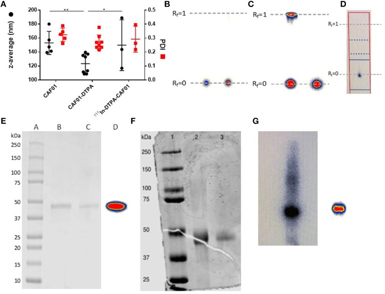
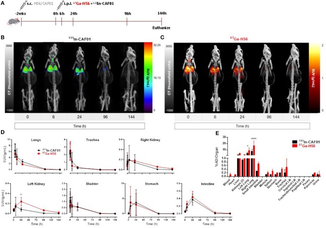
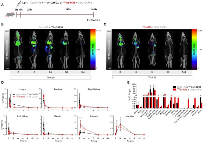
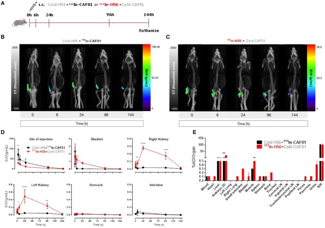
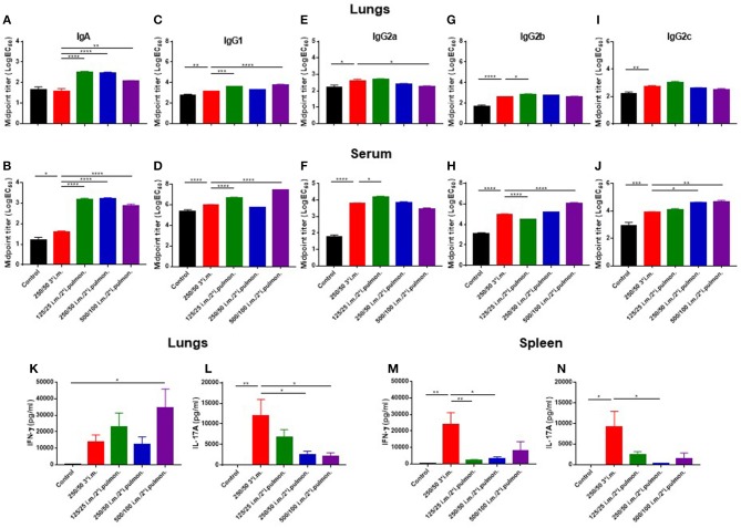
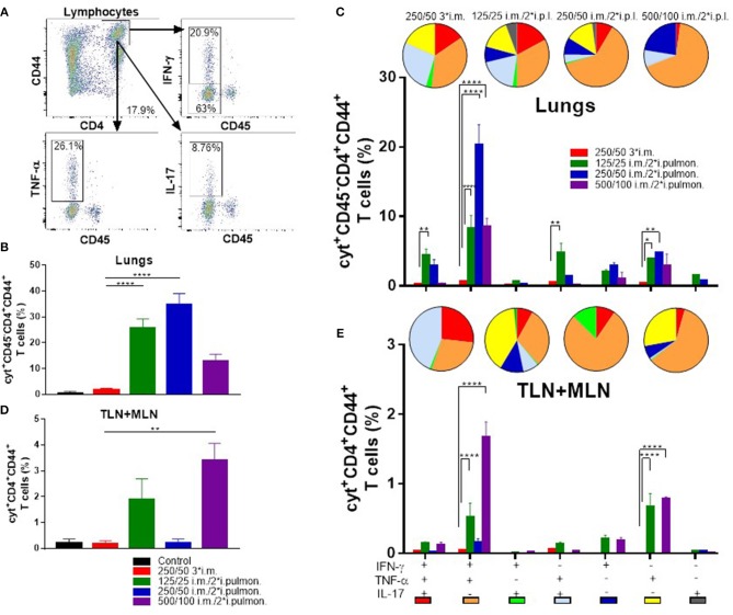
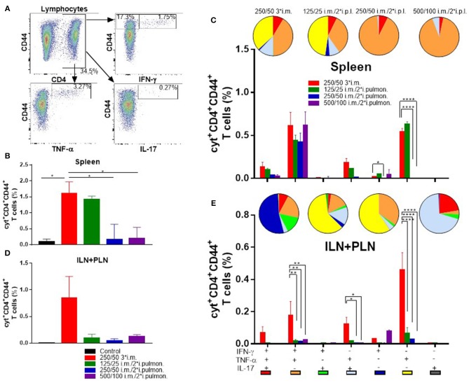
Pulmonary tuberculosis (TB), which is caused by (, remains a global pandemic, despite the widespread use of the parenteral live attenuated Bacillus Calmette-Guérin (BCG) vaccine during the past decades. Mucosal administration of next generation TB vaccines has great potential, but developing a safe and efficacious mucosal vaccine is challenging. Hence, understanding the biodistribution and pharmacokinetics of mucosal vaccines is essential for shaping the desired immune response and for optimal spatiotemporal targeting of the appropriate effector cells in the lungs. A subunit vaccine consisting of the fusion antigen H56 (Ag85B-ESAT-6-Rv2660) and the liposome-based cationic adjuvant formulation (CAF01) confers efficient protection in preclinical animal models. In this study, we devise a novel immunization strategy for the H56/CAF01 vaccine, which comply with the intrapulmonary (i.pulmon.) route of immunization. We also describe a novel dual-isotope (In/Ga) radiolabeling approach, which enables simultaneous non-invasive and longitudinal SPECT/CT imaging and quantification of H56 and CAF01 upon parenteral prime and/or i.pulmon. boost immunization. Our results demonstrate that the vaccine is distributed evenly in the lungs, and there are pronounced differences in the pharmacokinetics of H56 and CAF01. We provide convincing evidence that the H56/CAF01 vaccine is not only well-tolerated when administered to the respiratory tract, but it also induces strong lung mucosal and systemic IgA and polyfunctional Th1 and Th17 responses after parenteral prime and i.pulmon. boost immunization. The study furthermore evaluate the application of SPECT/CT imaging for the investigation of vaccine biodistribution after parenteral and i.pulmon. immunization of mice.
肺结核(TB),由 引起( ),尽管在过去几十年中广泛使用了皮下注射的减毒活卡介苗(BCG)疫苗,但仍在全球流行。下一代结核病疫苗的黏膜给药具有很大的潜力,但开发安全有效的黏膜疫苗具有挑战性。因此,了解黏膜疫苗的 生物分布和药代动力学对于塑造所需的免疫反应以及优化肺部适当效应细胞的时空靶向至关重要。由融合抗原 H56(Ag85B-ESAT-6-Rv2660)和基于脂质体的阳离子佐剂制剂(CAF01)组成的亚单位疫苗在临床前动物模型中提供了有效的保护。在这项研究中,我们设计了一种新的 H56/CAF01 疫苗免疫策略,符合肺内(i.pulmon.)免疫途径。我们还描述了一种新的双同位素(In/Ga)放射性标记方法,该方法能够在皮下注射初免和/或肺内加强免疫后,同时进行非侵入性和纵向 SPECT/CT 成像和定量检测 H56 和 CAF01。我们的结果表明,疫苗在肺部均匀分布,并且 H56 和 CAF01 的药代动力学存在明显差异。我们提供了令人信服的证据,证明 H56/CAF01 疫苗不仅在呼吸道给药时具有良好的耐受性,而且在皮下注射初免和肺内加强免疫后,还能诱导强烈的肺部黏膜和系统 IgA 以及多功能 Th1 和 Th17 反应。该研究还评估了 SPECT/CT 成像在研究皮下注射和肺内免疫后疫苗生物分布中的应用。