Lin Yun-Hsuan, Wang Nan-Kai, Yeung Ling, Lai Chi-Chun, Chuang Lan-Hsin
Department of Ophthalmology, Chang-Gung Memorial Hospital, 222 Mai-Chin Rd, Keelung, 204, Taiwan (Republic of China).
Department of Ophthalmology, Chang-Gung Memorial Hospital, Linkou, Taiwan.
BMC Ophthalmol. 2018 Dec 17;18(1):323. doi: 10.1186/s12886-018-0980-2.
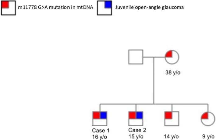
Leber's hereditary optic neuropathy (LHON) is a maternally inherited recessive disease rarely complicated with glaucoma. We conducted a clinical and genetic retrospective case series to describe three cases of juvenile open-angle glaucoma (JOAG) and an ND4 m11778G > A mitochondrial DNA (mtDNA) mutation, which is pathognomonic for LHON.
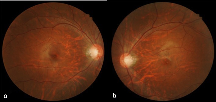
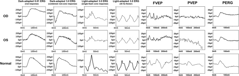
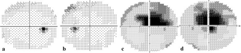
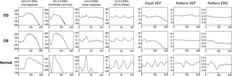
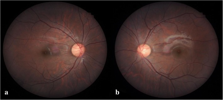
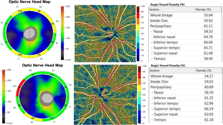
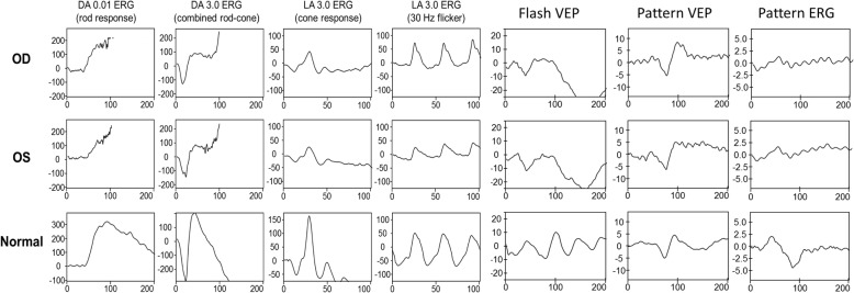
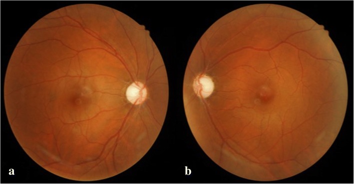
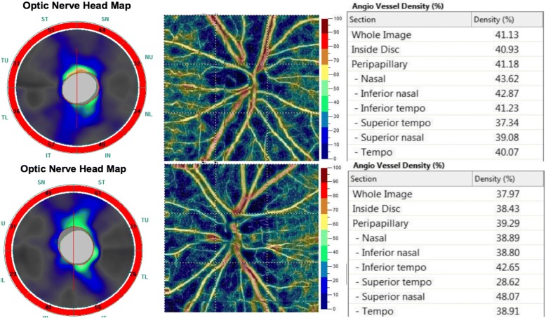
Patient 1 was a 16-year-old boy diagnosed with bilateral JOAG and high myopia. His intraocular pressure (IOP) was poorly controlled with the use of full topical anti-glaucoma medications. His best-corrected visual acuity (BCVA) decreased gradually over 5 years. Fundoscopic examination revealed bilateral enlarged disc cupping of the optic nerves with sectorial excavation and reduction of the neural rim in the left eye. His visual field (VF) was characterized by bilateral progressive central scotoma. Pattern visual evoked potentials (VEPs) and pattern electroretinograms (ERGs) showed extinguished responses in both eyes. Because of the non-specific visual field findings and the optic neuropathy disclosed by the pattern VEPs and pattern ERGs, we arranged a genetic test for the patient, which revealed an m11778G > A mtDNA mutation. Patient 2, the younger brother of Patient 1, was a 15-year-old boy who had been diagnosed with bilateral JOAG in 2010. The BCVA of both eyes remained at 1.0 during the follow-up period. Fundoscopic examination revealed bilateral mildly paled optic disc with enlarged cupping and reduction of the neural rim. The pattern ERG revealed a decreased N95 amplitude bilaterally. The genetic test revealed an m11778G > A mtDNA mutation. Patient 3 was a 35-year-old man with bilateral JOAG. His BCVA decreased gradually over 10 years. Fundoscopic examination revealed paled optic disc with enlarged disc cupping and reduction of the neural rim in both eyes. The pattern ERG revealed a decreased N95 amplitude bilaterally. The genetic test revealed an m11778G > A mtDNA mutation.
This case series describes three patients with concomitant occurrence of JOAG and LHON. These two diseases may have a cumulative effect on oxidative stress and retinal ganglion cell death with the rapid deterioration of vision, which may occur during adolescence.
Leber遗传性视神经病变(LHON)是一种母系遗传的隐性疾病,很少并发青光眼。我们进行了一项临床和遗传学回顾性病例系列研究,以描述3例青少年开角型青光眼(JOAG)患者及11778G>A线粒体DNA(mtDNA)突变,该突变是LHON的特征性表现。
患者1为一名16岁男孩,诊断为双眼JOAG及高度近视。使用所有局部抗青光眼药物后,其眼压控制不佳。其最佳矫正视力(BCVA)在5年中逐渐下降。眼底检查显示双侧视神经盘杯状扩大,左眼有扇形凹陷及神经边缘减少。其视野(VF)表现为双侧进行性中心暗点。图形视觉诱发电位(VEP)和图形视网膜电图(ERG)显示双眼反应消失。由于视野检查结果不具特异性,且图形VEP和图形ERG显示存在视神经病变,我们为该患者安排了基因检测,结果显示存在m11778G>A mtDNA突变。患者2是患者1的弟弟,为一名15岁男孩,于2010年被诊断为双眼JOAG。随访期间双眼BCVA保持在1.0。眼底检查显示双侧视神经盘轻度苍白,杯状扩大,神经边缘减少。图形ERG显示双侧N95波幅降低。基因检测显示存在m11778G>A mtDNA突变。患者3为一名35岁男性,患有双眼JOAG。其BCVA在10年中逐渐下降。眼底检查显示双眼视神经盘苍白,盘杯状扩大,神经边缘减少。图形ERG显示双侧N95波幅降低。基因检测显示存在m11778G>A mtDNA突变。
该病例系列描述了3例同时患有JOAG和LHON的患者。这两种疾病可能对氧化应激和视网膜神经节细胞死亡产生累积效应,导致视力迅速恶化,这种情况可能在青春期发生。