Dastoori Mahmoud, Bouserhal Joseph P, Halazonetis Demetrios J, Athanasiou Athanasios E
Former resident, Department of Orthodontics, Hamdan Bin Mohammed College of Dental Medicine, Mohammed Bin Rashid University of Medicine and Health Sciences, Dubai, UAE; Orthodontic Specialist, London Dental Clinic, Dubai, UAE.
Professor, Department of Orthodontics, Faculty of Dental Medicine, Saint Joseph University of Beirut, Beirut, Lebanon.
J Clin Exp Dent. 2018 Nov 1;10(11):e1069-e1074. doi: 10.4317/jced.55180. eCollection 2018 Nov.
To assess the accuracy of digital models generated using commercially available software to predict anterior teeth root inclination characteristics and compare the results to relevant data obtained from CBCT images.
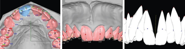
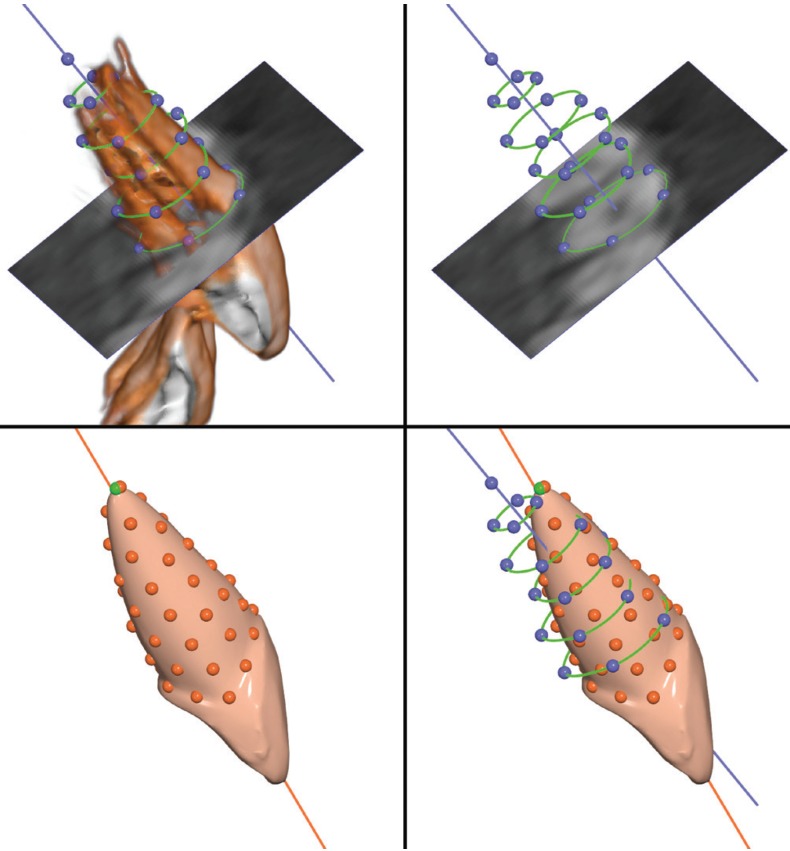
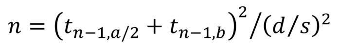
Following sample size calculation and after application of inclusion and exclusion criteria, pre-treatment maxillary and mandibular plaster models and the corresponding CBCT scans of 31 patients attending a private orthodontic clinic were selected. The subjects were 10 males and 21 females with age range 12 to 40 years. Plaster models were scanned using the high resolution mode of an Ortho Insight 3D scanner and CBCT scans were taken using a Kodak 9500 Cone Beam 3D System machine. The teeth on the digital scans were segmented and virtual roots were predicted and constructed by the Ortho Insight 3D software. The long axes of the predicted roots and the actual roots, as segmented from the CBCT images, were computed using best-fit lines. The inter-axis angle was used to assess error in root inclination prediction by the software. Mann-Whitney and Kruskal-Wallis tests were used. Intra-examiner error was evaluated using the Bland-Altman method.
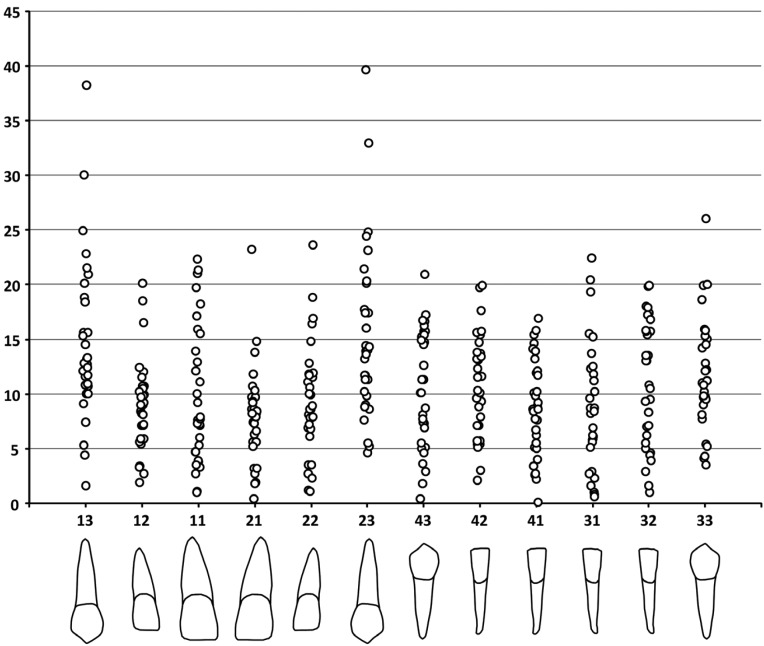
The maximum disparity in angle between images derived from digital models and CBCT data was almost 40 degrees (upper left canine). The upper and lower canines produced the worst results, followed by the lower lateral incisors. The upper central incisors showed the best results, although the maximum angle of difference exceeded 20 degrees (with the median around 8 degrees).
Root morphology imaging prediction is not a primary function of this software and this study confirmed its limitation as a sole tool in routine clinical applications. At present these predictions cannot be considered accurate or reliable unless correlated clinically with a radiographic image. Digital models, CBCT, tooth root inclination prediction software.
评估使用商用软件生成的数字模型预测前牙牙根倾斜特征的准确性,并将结果与从CBCT图像获得的相关数据进行比较。
在计算样本量并应用纳入和排除标准后,选取了一家私立正畸诊所31例患者的治疗前上颌和下颌石膏模型以及相应的CBCT扫描图像。受试者为10名男性和21名女性,年龄在12至40岁之间。使用Ortho Insight 3D扫描仪的高分辨率模式扫描石膏模型,并使用柯达9500锥形束3D系统机器进行CBCT扫描。对数字扫描图像上的牙齿进行分割,并通过Ortho Insight 3D软件预测和构建虚拟牙根。使用最佳拟合线计算预测牙根和从CBCT图像分割出的实际牙根的长轴。轴间角度用于评估软件预测牙根倾斜的误差。采用曼-惠特尼检验和克鲁斯卡尔-沃利斯检验。使用布兰德-奥特曼方法评估检查者内部误差。
数字模型图像与CBCT数据之间的最大角度差异接近40度(左上尖牙)。上下尖牙的结果最差,其次是下颌侧切牙。上颌中切牙的结果最好,尽管最大差异角度超过20度(中位数约为8度)。
牙根形态成像预测不是该软件的主要功能,本研究证实了其作为常规临床应用中唯一工具的局限性。目前,除非在临床上与放射图像相关联,否则这些预测不能被认为是准确或可靠的。数字模型、CBCT、牙根倾斜预测软件。