Shi Lei, Cavagnino Andrea, Rabefiraisana Jean-Luc, Lazar Noureddine, Li de la Sierra-Gallay Inès, Ochsenbein Françoise, Valerio-Lepiniec Marie, Urvoas Agathe, Minard Philippe, Mijakovic Ivan, Nessler Sylvie
Division of Systems and Synthetic Biology, Department of Chemical and Biological Engineering, Chalmers University of Technology, Gothenburg, Sweden.
Institute of Integrative Biology of the Cell (I2BC), CEA, CNRS, Université Paris-Sud, Université Paris-Saclay, Gif-sur-Yvette, France.
Front Microbiol. 2019 Jan 8;9:3014. doi: 10.3389/fmicb.2018.03014. eCollection 2018.
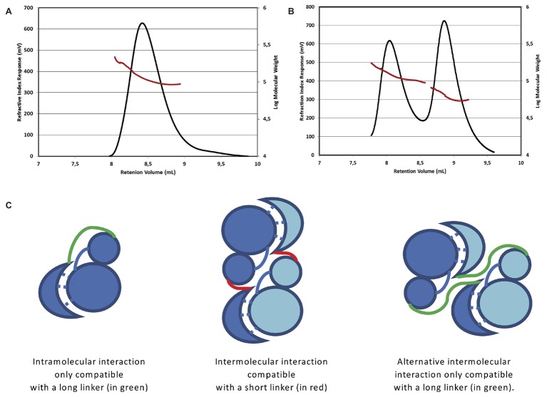
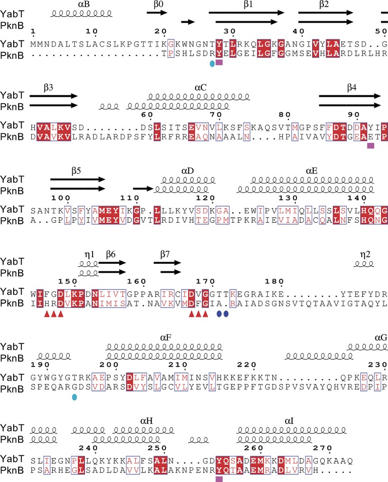
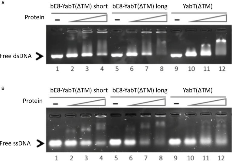
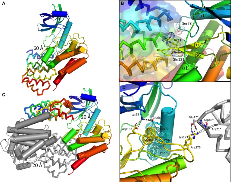
YabT is a serine/threonine kinase of the Hanks family from , which lacks the canonical extracellular signal receptor domain but is anchored to the membrane through a C-terminal transmembrane helix. A previous study demonstrated that a basic juxtamembrane region corresponds to a DNA-binding motif essential for the activation of YabT trans-autophosphorylation. YabT is expressed during spore development and localizes to the asymmetric septum where it specifically phosphorylates essential proteins involved in genome maintenance, such as RecA, SsbA, and YabA. YabT has also been shown to phosphorylate proteins involved in protein synthesis, such as AbrB and Ef-Tu, suggesting a possible regulatory role in the progressive metabolic quiescence of the forespore. Finally, cross phosphorylations with other protein kinases implicate YabT in the regulation of numerous other cellular processes. Using an artificial protein scaffold as crystallization helper, we determined the first crystal structure of this DNA-dependent bacterial protein kinase. This allowed us to trap the active conformation of the kinase domain of YabT. Using NMR, we showed that the basic juxtamembrane region of YabT is disordered in the absence of DNA in solution, just like it is in the crystal, and that it is stabilized upon DNA binding. In comparison with its closest structural homolog, the mycobacterial kinase PknB allowed us to discuss the dimerization mode of YabT. Together with phosphorylation assays and DNA-binding experiments, this structural analysis helped us to gain new insights into the regulatory activation mechanism of YabT.
YabT是一种来自[具体来源未提及]的Hanks家族丝氨酸/苏氨酸激酶,它缺乏典型的细胞外信号受体结构域,但通过C末端跨膜螺旋锚定在膜上。先前的一项研究表明,一个碱性近膜区域对应于激活YabT自磷酸化所必需的DNA结合基序。YabT在孢子发育过程中表达,并定位于不对称隔膜,在那里它特异性地磷酸化参与基因组维持的必需蛋白质,如RecA、SsbA和YabA。YabT也已被证明可磷酸化参与蛋白质合成的蛋白质,如AbrB和Ef-Tu,这表明它在芽孢前体的渐进性代谢静止中可能具有调节作用。最后,与其他蛋白激酶的交叉磷酸化表明YabT参与了许多其他细胞过程的调节。我们使用人工蛋白质支架作为结晶辅助物,确定了这种DNA依赖性细菌蛋白激酶的首个晶体结构。这使我们能够捕获YabT激酶结构域的活性构象。通过核磁共振,我们表明YabT的碱性近膜区域在溶液中不存在DNA时是无序的,就像在晶体中一样,并且在DNA结合时会稳定下来。与其最接近的结构同源物——分枝杆菌激酶PknB相比,这使我们能够讨论YabT的二聚化模式。结合磷酸化测定和DNA结合实验,这种结构分析帮助我们对YabT的调节激活机制有了新的认识。