The Hatter Cardiovascular Institute, University College London, 67 Chenies Mews, London WC1E 6HX, UK.
The Hatter Cardiovascular Institute, University College London, 67 Chenies Mews, London WC1E 6HX, UK.
J Mol Cell Cardiol. 2019 Mar;128:187-197. doi: 10.1016/j.yjmcc.2019.02.002. Epub 2019 Feb 7.
The chemokine stromal derived factor-1α (SDF-1α) is known to protect the heart acutely from ischaemia-reperfusion injury via its cognate receptor, CXCR4. However, the timing and cellular location of this effect, remains controversial.
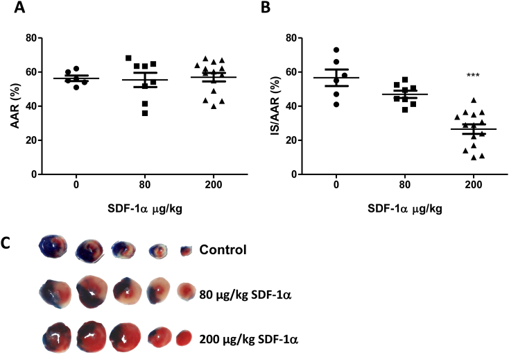
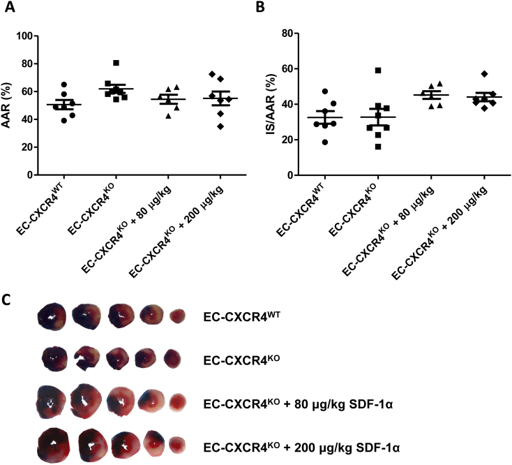
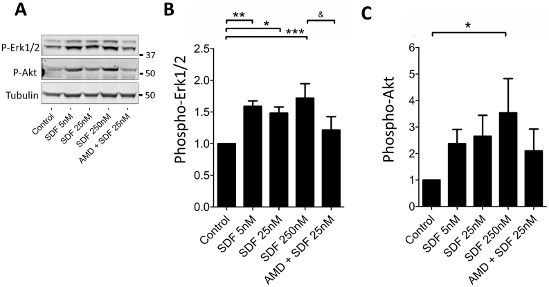
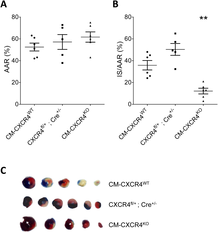
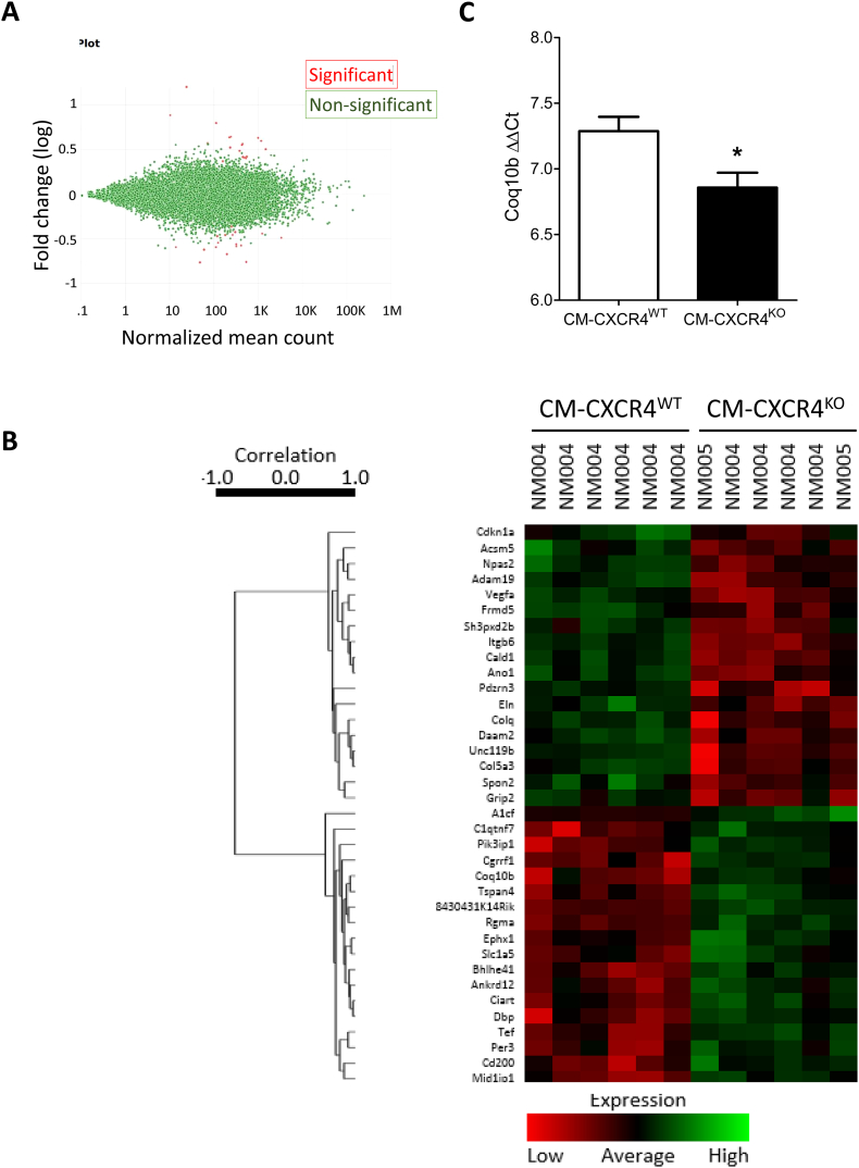
Wild type male and female mice were subjected to 40 min LAD territory ischaemia in vivo and injected with either saline (control) or SDF-1α prior to 2 h reperfusion. Infarct size as a proportion of area at risk was assessed histologically using Evans blue and triphenyltetrazolium chloride. Our results confirm the cardioprotective effect of exogenous SDF-1α in mouse ischaemia-reperfusion injury and, for the first time, show protection when SDF-1α is delivered just prior to reperfusion, which has important therapeutic implications. The role of cell type was examined using the same in vivo ischaemia-reperfusion protocol in cardiomyocyte- and endothelial-specific CXCR4-null mice, and by Western blot analysis of endothelial cells treated in vitro. These experiments demonstrated that the acute infarct-sparing effect is mediated by endothelial cells, possibly via the signalling kinases Erk1/2 and PI3K/Akt. Unexpectedly, cardiomyocyte-specific deletion of CXCR4 was found to be cardioprotective per se. RNAseq analysis indicated altered expression of the mitochondrial protein co-enzyme Q10b in these mice.
Administration of SDF-1α is cardioprotective when administered prior to reperfusion and may, therefore, have clinical utility. SDF-1α-CXCR4-mediated cardioprotection from ischaemia-reperfusion injury is contingent on the cellular location of CXCR4 activation. Specifically, cardioprotection is mediated by endothelial signalling, while cardiomyocyte-specific deletion of CXCR4 has an infarct-sparing effect per se.
趋化因子基质衍生因子-1α(SDF-1α)通过其同源受体 CXCR4,已知可在急性缺血再灌注损伤中保护心脏。然而,这种效应的时间和细胞位置仍存在争议。
体内雄性和雌性野生型小鼠接受 40 分钟左前降支(LAD)区域缺血,并在再灌注前 2 小时注射盐水(对照)或 SDF-1α。使用 Evans 蓝和三苯基四唑氯盐通过组织学评估梗塞面积占危险区域的比例。我们的结果证实了外源性 SDF-1α 在小鼠缺血再灌注损伤中的心脏保护作用,并且首次显示 SDF-1α 在再灌注前给药时具有保护作用,这具有重要的治疗意义。使用相同的体内缺血再灌注方案在心肌细胞和内皮细胞特异性 CXCR4 缺失小鼠中检查细胞类型的作用,并通过体外处理的内皮细胞的 Western blot 分析。这些实验表明,急性梗塞保护作用是由内皮细胞介导的,可能通过信号激酶 Erk1/2 和 PI3K/Akt 介导。出乎意料的是,发现心肌细胞特异性缺失 CXCR4 本身具有心脏保护作用。RNAseq 分析表明,这些小鼠中线粒体蛋白辅酶 Q10b 的表达发生改变。
在再灌注前给予 SDF-1α 是心脏保护的,因此可能具有临床效用。SDF-1α-CXCR4 介导的缺血再灌注损伤的心脏保护取决于 CXCR4 激活的细胞位置。具体而言,内皮细胞信号介导心脏保护,而心肌细胞特异性缺失 CXCR4 本身具有梗塞保护作用。