• 文献检索
  • 文档翻译
  • 深度研究
  • 学术资讯
  • Suppr Zotero 插件Zotero 插件
  • 邀请有礼
  • 套餐&价格
  • 历史记录
应用&插件
Suppr Zotero 插件Zotero 插件浏览器插件Mac 客户端Windows 客户端微信小程序
定价
高级版会员购买积分包购买API积分包
服务
文献检索文档翻译深度研究API 文档MCP 服务
关于我们
关于 Suppr公司介绍联系我们用户协议隐私条款
关注我们

Suppr 超能文献

核心技术专利:CN118964589B侵权必究
粤ICP备2023148730 号-1Suppr @ 2026

文献检索

告别复杂PubMed语法,用中文像聊天一样搜索,搜遍4000万医学文献。AI智能推荐,让科研检索更轻松。

立即免费搜索

文件翻译

保留排版,准确专业,支持PDF/Word/PPT等文件格式,支持 12+语言互译。

免费翻译文档

深度研究

AI帮你快速写综述,25分钟生成高质量综述,智能提取关键信息,辅助科研写作。

立即免费体验

恒牙上颌第一磨牙近中颊根第二根管的几何特征:一项锥形束计算机断层扫描研究

Geometric features of second mesiobuccal canal in permanent maxillary first molars: a cone-beam computed tomography study.

作者信息

Su Chi-Chun, Wu Yu-Chiao, Chung Ming-Pang, Huang Ren-Yeong, Cheng Wan-Chien, Cathy Tsai Yi-Wen, Hsieh Chen-Yu, Chiang Ho-Sheng, Chen Ching-Yang, Shieh Yi-Shing

机构信息

School of Dentistry, Tri-Service General Hospital and National Defense Medical Center, Taipei, Taiwan.

Radiology Division, SongShan Branch, Tri-Service General Hospital and National Defense Medical Center, Taipei, Taiwan.

出版信息

J Dent Sci. 2017 Sep;12(3):241-248. doi: 10.1016/j.jds.2017.03.002. Epub 2017 Apr 2.

DOI:10.1016/j.jds.2017.03.002
PMID:30895057
原文链接:https://pmc.ncbi.nlm.nih.gov/articles/PMC6400017/
Abstract

BACKGROUND/PURPOSE: Inadequacy to locate the second mesiobuccal canal (MB2) canal leads to the highest probability of endodontic failure in permanent maxillary first molars (PMFMs) and still remains a constant challenge for many clinicians. The aim of this study was to characterize the geometrical features between MB2 and other orifices of examined PMFMs using cone-beam computed tomography images.

MATERIALS AND METHODS

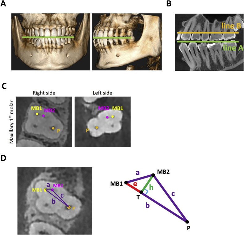
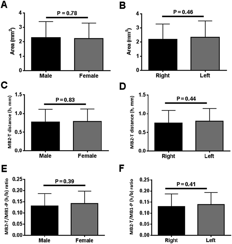
A total of 108 teeth qualified in the cone-beam computed tomography image archives were enrolled in the present study. The intersecting point (T) was determined as the perpendicular line (h, altitude of triangle) projected from the vertex of the MB2 canal orifice to mesiobuccal canal orifice-palatal canal orifice line (MB1-P). We measured the geometric features of PMFMs with the MB2 canal, including the interorifice distances, area, altitude, and the ratio between the canal orifices.

RESULTS

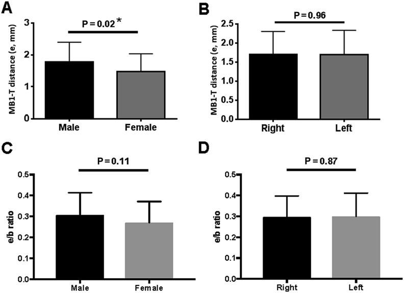
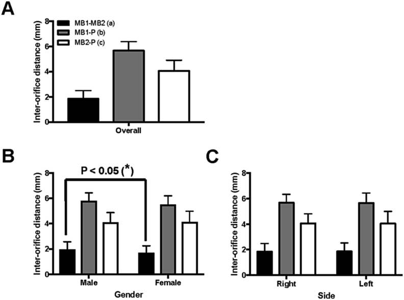
The average interorifice distance was found to be 1.91 ± 0.59 mm for MB1-MB2, 5.73 ± 0.66 mm for MB1-P, and 4.11 ± 0.79 mm for MB2-P, with significant gender difference for MB1-MB2 distance. For the MB1-T distance, a significant difference was found between genders (P = 0.02), with males averaging 1.78 ± 0.07 mm, and females 1.48 ± 0.11 mm. For the MB1-P distance, the majorities of both genders were found in the 20-40% cut-off. A portion of the males exhibited a tendency towards the 40-80% cut-off, while females shifted in the reverse direction towards the 0-20% cut-off.

CONCLUSION

In clinical scenarios, these anatomical characteristics of the root canals system could be beneficial to locating the MB2 canal.

摘要

背景/目的:难以找到上颌第一恒磨牙(PMFMs)的第二近中颊根管(MB2)会导致根管治疗失败的可能性最高,这对许多临床医生来说仍是一项持续的挑战。本研究的目的是使用锥形束计算机断层扫描图像来描述所检查的PMFMs中MB2与其他根管口之间的几何特征。

材料与方法

本研究共纳入了108颗符合锥形束计算机断层扫描图像存档要求的牙齿。相交点(T)被确定为从MB2根管口顶点向近中颊根管口-腭根管口连线(MB1-P)投射的垂线(h,三角形的高)。我们测量了具有MB2根管的PMFMs的几何特征,包括根管口间距离、面积、高度以及根管口之间的比例。

结果

发现MB1-MB2的平均根管口间距离为1.91±0.59毫米,MB1-P为5.73±0.66毫米,MB2-P为4.11±0.79毫米,MB1-MB2距离存在显著的性别差异。对于MB1-T距离,性别之间存在显著差异(P = 0.02),男性平均为1.78±0.07毫米,女性为1.48±0.11毫米。对于MB1-P距离,大多数男性和女性都处于20%-40%的区间。一部分男性有向40%-80%区间偏移的趋势,而女性则向相反方向偏移至0%-20%区间。

结论

在临床情况下,根管系统的这些解剖特征可能有助于定位MB2根管。

https://cdn.ncbi.nlm.nih.gov/pmc/blobs/adc3/6400017/d8f238a19bae/gr4.jpg
https://cdn.ncbi.nlm.nih.gov/pmc/blobs/adc3/6400017/20ba0aee5675/gr1.jpg
https://cdn.ncbi.nlm.nih.gov/pmc/blobs/adc3/6400017/329eaffb14d5/gr2.jpg
https://cdn.ncbi.nlm.nih.gov/pmc/blobs/adc3/6400017/06cd1be33b45/gr3.jpg
https://cdn.ncbi.nlm.nih.gov/pmc/blobs/adc3/6400017/d8f238a19bae/gr4.jpg
https://cdn.ncbi.nlm.nih.gov/pmc/blobs/adc3/6400017/20ba0aee5675/gr1.jpg
https://cdn.ncbi.nlm.nih.gov/pmc/blobs/adc3/6400017/329eaffb14d5/gr2.jpg
https://cdn.ncbi.nlm.nih.gov/pmc/blobs/adc3/6400017/06cd1be33b45/gr3.jpg
https://cdn.ncbi.nlm.nih.gov/pmc/blobs/adc3/6400017/d8f238a19bae/gr4.jpg

相似文献

1
Geometric features of second mesiobuccal canal in permanent maxillary first molars: a cone-beam computed tomography study.恒牙上颌第一磨牙近中颊根第二根管的几何特征:一项锥形束计算机断层扫描研究
J Dent Sci. 2017 Sep;12(3):241-248. doi: 10.1016/j.jds.2017.03.002. Epub 2017 Apr 2.
2
Assessment of the Second Mesiobuccal Root Canal in Maxillary First Molars: A Cone-beam Computed Tomographic Study.上颌第一磨牙第二近中颊根根管评估:锥形束 CT 研究。
J Endod. 2017 Dec;43(12):1990-1996. doi: 10.1016/j.joen.2017.06.021. Epub 2017 Oct 9.
3
Assessment of the presence of a second mesiobuccal canal in maxillary first molars according to the location of the main mesiobuccal canal-a micro-computed tomographic study.上颌第一磨牙第二近中颊根管存在情况的评估:基于主近中颊根管位置的分析-一项显微 CT 研究。
Clin Oral Investig. 2021 Jun;25(6):3937-3944. doi: 10.1007/s00784-020-03723-5. Epub 2021 Jan 6.
4
A cone-beam computed tomography study of the prevalence and location of the second mesiobuccal root canal in maxillary molars.一项关于上颌磨牙近中颊侧第二根管患病率及位置的锥形束计算机断层扫描研究。
Restor Dent Endod. 2020 Sep 3;45(4):e46. doi: 10.5395/rde.2020.45.e46. eCollection 2020 Nov.
5
Detection and location of second mesiobuccal canal in permanent maxillary teeth: A cone-beam computed tomography analysis in a Taiwanese population.在台湾人群中,通过锥形束 CT 分析发现上颌恒磨牙的第二近中颊根管:一项研究。
Arch Oral Biol. 2019 Feb;98:108-114. doi: 10.1016/j.archoralbio.2018.11.006. Epub 2018 Nov 12.
6
Location of the Second Mesiobuccal Canal of Maxillary Molars in a Brazilian Subpopulation: Analyzing Using Cone-beam Computed Tomography.上颌磨牙第二近颊管在巴西亚群中的位置:锥形束 CT 分析。
J Contemp Dent Pract. 2022 Oct 1;23(10):979-983. doi: 10.5005/jp-journals-10024-3422.
7
Cone-beam computed tomography study of prevalence and location of MB2 canal in the mesiobuccal root of the maxillary second molar.上颌第二磨牙近中颊根MB2根管患病率及位置的锥形束计算机断层扫描研究
Int J Clin Exp Med. 2015 Jun 15;8(6):9128-34. eCollection 2015.
8
Micro-CT evaluation of maxillary first molars: interorifice distances and internal anatomy of the mesiobuccal root.上颌第一磨牙的显微CT评估:近中颊根的根管口间距及内部解剖结构
Braz Oral Res. 2021 Apr 26;35:e060. doi: 10.1590/1807-3107bor-2021.vol35.0060. eCollection 2021.
9
Cone-beam computed tomography study of the incidence and characteristics of the second mesiobuccal canal in maxillary permanent molars.上颌恒磨牙近中颊根第二根管发生率及特征的锥形束计算机断层扫描研究
Front Physiol. 2022 Nov 24;13:993006. doi: 10.3389/fphys.2022.993006. eCollection 2022.
10
Mesiobuccal and Palatal Interorifice Distance May Predict the Presence of the Second Mesiobuccal Canal in Maxillary Second Molars with Fused Roots.颊腭向根管间距离可预测融合根上颌第二磨牙第二近中颊根管的存在。
J Endod. 2021 Apr;47(4):585-591. doi: 10.1016/j.joen.2021.01.003. Epub 2021 Jan 23.

引用本文的文献

1
An Evaluation of the Relationship Between the Mesiobuccal Canal Configuration, the Interorifice Distance, and the Root Lengths of the Permanent Maxillary First Molars with Cone Beam Computed Tomography.锥形束计算机断层扫描评估上颌第一恒磨牙近中颊根管形态、根管口间距与牙根长度之间的关系
Diagnostics (Basel). 2024 Nov 30;14(23):2703. doi: 10.3390/diagnostics14232703.
2
Apical microsurgery of C-shaped maxillary first molar: A case report.C形上颌第一磨牙根尖显微手术:一例报告
J Dent Sci. 2021 Jul;16(3):1035-1036. doi: 10.1016/j.jds.2020.11.003. Epub 2020 Nov 23.
3
Root canal isthmi and interorifice distance in human permanent teeth of an Indian subpopulation using cone-beam computed tomography: A retrospective cross-sectional study.

本文引用的文献

1
CBCT uses in clinical endodontics: the effect of CBCT on the ability to locate MB2 canals in maxillary molars.CBCT 在临床牙髓学中的应用:CBCT 对上颌磨牙 MB2 根管定位能力的影响。
Int Endod J. 2017 Dec;50(12):1109-1115. doi: 10.1111/iej.12736. Epub 2017 Jan 17.
2
Prevalence and location of the secondary mesiobuccal canal in 1,100 maxillary molars using cone beam computed tomography.使用锥形束计算机断层扫描技术对1100颗上颌磨牙中第二近中颊根管的患病率及位置进行研究。
BMC Med Imaging. 2016 Dec 1;16(1):66. doi: 10.1186/s12880-016-0168-2.
3
Analysis of the Root and Canal Morphologies in Maxillary First and Second Molars in a Chinese Population Using Cone-beam Computed Tomography.
使用锥形束计算机断层扫描对印度亚人群恒牙根管峡部和根管口间距离的研究:一项回顾性横断面研究
J Conserv Dent. 2020 Nov-Dec;23(6):563-570. doi: 10.4103/JCD.JCD_576_20. Epub 2021 Feb 11.
4
Assessment of Mesiobuccal Canal Configuration, Prevalence and Inter-Orifice Distance at Different Root Thirds of Maxillary First Molars: A CBCT Study.上颌第一磨牙不同牙根三分之一处近中颊根管形态、发生率及根管口间距的评估:一项锥形束CT研究
Clin Cosmet Investig Dent. 2021 Mar 24;13:105-111. doi: 10.2147/CCIDE.S300241. eCollection 2021.
5
Three-Dimensional Cone-Beam Computed Tomography Assessment of Additional Canals of Permanent first Molars: A Pinocchio for Successful Root Canal Treatment.三维锥形束计算机断层扫描评估恒牙第一磨牙的额外根管:成功进行根管治疗的关键因素
J Int Soc Prev Community Dent. 2018 May-Jun;8(3):259-263. doi: 10.4103/jispcd.JISPCD_3_18. Epub 2018 May 17.
使用锥形束计算机断层扫描分析中国人群上颌第一和第二磨牙的根管形态
J Endod. 2016 May;42(5):696-701. doi: 10.1016/j.joen.2016.01.017. Epub 2016 Mar 16.
4
Preoperative assessment of labial bone perforation for virtual immediate implant surgery in the maxillary esthetic zone: A computer simulation study.上颌美学区虚拟即刻种植手术唇侧骨穿孔的术前评估:一项计算机模拟研究。
J Am Dent Assoc. 2015 Nov;146(11):808-19. doi: 10.1016/j.adaj.2015.04.007.
5
Cone-beam computed tomography study of prevalence and location of MB2 canal in the mesiobuccal root of the maxillary second molar.上颌第二磨牙近中颊根MB2根管患病率及位置的锥形束计算机断层扫描研究
Int J Clin Exp Med. 2015 Jun 15;8(6):9128-34. eCollection 2015.
6
Accuracy of Cone-beam Computed Tomography in the Detection of a Second Mesiobuccal Root Canal in Endodontically Treated Teeth: An Ex Vivo Study.锥形束计算机断层扫描在检测根管治疗牙齿中第二近中颊根管的准确性:一项体外研究
J Endod. 2015 Oct;41(10):1678-81. doi: 10.1016/j.joen.2015.06.011. Epub 2015 Aug 15.
7
The Diagnostic Efficacy of Cone-beam Computed Tomography in Endodontics: A Systematic Review and Analysis by a Hierarchical Model of Efficacy.锥形束计算机断层扫描在牙髓病学中的诊断效能:基于效能分层模型的系统评价与分析
J Endod. 2015 Jul;41(7):1008-14. doi: 10.1016/j.joen.2015.02.021. Epub 2015 Apr 3.
8
Frequency of second mesiobuccal canal in permanent maxillary first molars using the operating microscope and selective dentin removal: A clinical study.使用手术显微镜和选择性牙本质去除术观察上颌第一恒磨牙近中颊根第二根管的发生率:一项临床研究
Contemp Clin Dent. 2015 Jan-Mar;6(1):74-8. doi: 10.4103/0976-237X.149296.
9
Determination of frequency of the second mesiobuccal canal in the permanent maxillary first molar teeth with magnification loupes (× 3.5).使用放大倍数为3.5倍的放大镜确定恒牙上颌第一磨牙中第二近中颊根管的频率。
Int J Biomed Sci. 2014 Sep;10(3):201-7.
10
Evaluation of root and canal morphology of maxillary permanent first molars in a North American population by cone-beam computed tomography.通过锥形束计算机断层扫描评估北美人群上颌第一恒磨牙的根管形态。
J Endod. 2014 May;40(5):635-9. doi: 10.1016/j.joen.2014.02.002. Epub 2014 Mar 29.