Division of Structural Biology, Wellcome Trust Centre for Human Genetics, University of Oxford, Oxford, OX3 7BN, UK; Electron Bio-Imaging Centre, Diamond Light Source, Harwell Science and Innovation Campus, Didcot OX11 0DE, UK; Department of Structural Biology, University of Pittsburgh School of Medicine, Pittsburgh, PA 15260, USA.
Curr Opin Struct Biol. 2019 Oct;58:249-258. doi: 10.1016/j.sbi.2019.05.021. Epub 2019 Jul 5.
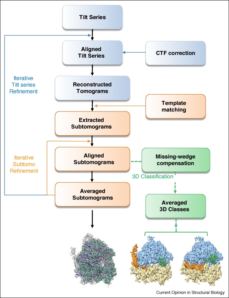
Cryo-electron tomography (cryoET) can provide 3D reconstructions, or tomograms, of pleomorphic objects such as organelles or cells in their close-to-native states. Subtomograms that contain repetitive structures can be further extracted and subjected to averaging and classification to improve resolution, and this process has become an emerging structural biology method referred to as cryoET subtomogram averaging and classification (cryoSTAC). Recent technical advances in cryoSTAC have had a profound impact on many fields in biology. Here, I review recent exciting work on several macromolecular assemblies demonstrating the power of cryoSTAC for in situ structure analysis and discuss challenges and future directions.
低温电子断层扫描(cryoET)可以提供类似原生状态下的细胞器或细胞等多形物体的 3D 重建,或断层扫描图像。可以进一步提取包含重复结构的亚断层扫描图像,并进行平均和分类,以提高分辨率,这个过程已成为一种新兴的结构生物学方法,称为 cryoET 亚断层扫描图像平均和分类(cryoSTAC)。cryoSTAC 的最新技术进展对生物学的许多领域都产生了深远的影响。在这里,我回顾了 cryoSTAC 在几个大分子组装体方面的令人兴奋的最新工作,展示了 cryoSTAC 用于原位结构分析的强大功能,并讨论了挑战和未来方向。