Department of Orthopaedics, Sun Yat-sen Memorial Hospital, Sun Yat-sen University, NO.107, Western Yanjiang Road, Yuexiu district, Guangzhou, Guangdong Province, 510120, People's Republic of China.
BMC Musculoskelet Disord. 2019 Jul 29;20(1):350. doi: 10.1186/s12891-019-2696-8.
Isolated rectus femoris (RF) contracture is encountered very rarely in orthopaedic practices. There are few reports on its imaging manifestations and no cases reported to be treated with arthroscopy.
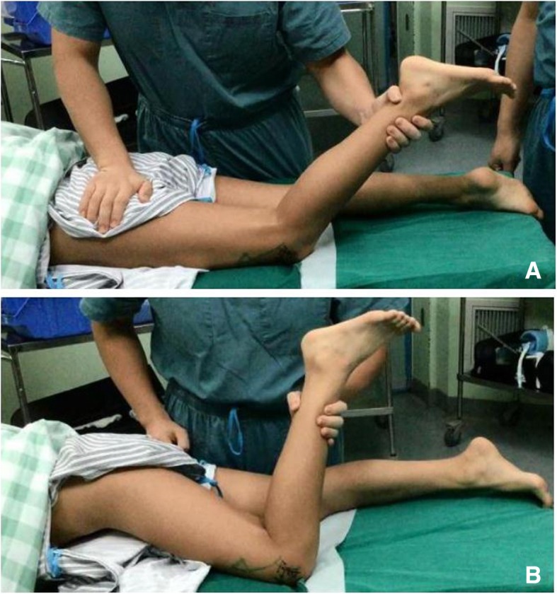
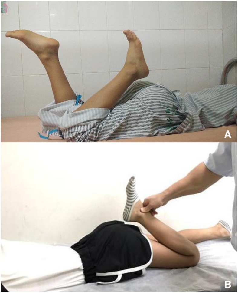
A 11-year-old girl with a more than 7 years history of restricted left knee flexion was presented. The clinical assessment and magnetic resonance imaging (MRI) findings were detailed here. A strip-like induration was palpated in the left thigh, which tends to be more obvious with knee flexion. MRI demonstrated a hypointensity band connected the anterior inferior iliac spine with the patella, and marked atrophy of the left RF muscle. Fibrosis contracture band was confirmed with arthroscope, then divided by radiofrequency ablation (RFA) under arthroscopic observation. Followed by debridement of the fibrillar connective tissue and hemostasis around the broken ends. The movement of left knee joint significantly improved after the operation, and the patient recovered nearly full range of motion of this joint after 6 months.
The specific MRI findings could assist in confirming clinical early diagnosis of isolated RF contracture. Arthroscopic RFA treatment is an effective technique to treat this disorder with minimally incision.
孤立性股直肌(RF)挛缩在骨科实践中非常罕见。关于其影像学表现的报道很少,也没有报道通过关节镜治疗的病例。
一名 11 岁女孩,左膝关节屈曲受限超过 7 年。详细介绍了临床评估和磁共振成像(MRI)的结果。左大腿可触及一条带状硬结,在膝关节屈曲时更为明显。MRI 显示一条连接髂前下棘和髌骨的低信号带,左 RF 肌肉明显萎缩。关节镜下证实为纤维性挛缩带,然后在关节镜观察下通过射频消融(RFA)进行分割。随后对纤维状结缔组织和断端周围的出血进行清创。术后左膝关节活动明显改善,6 个月后患者恢复了该关节的几乎全部活动范围。
特定的 MRI 发现有助于确认孤立性 RF 挛缩的临床早期诊断。关节镜下 RFA 治疗是一种微创治疗该疾病的有效技术。