Department of Radiology, Linköping University, Linköping, Sweden.
Department of Medical & Health Sciences, Linköping University, Linköping, Sweden.
BMC Med Imaging. 2019 Aug 9;19(1):64. doi: 10.1186/s12880-019-0363-z.
Our aim was to compare CT images from native, nephrographic and excretory phases using image quality criteria as well as the detection of positive pathological findings in CT Urography, to explore if the radiation burden to the younger group of patients or patients with negative outcomes can be reduced.
This is a retrospective study of 40 patients who underwent a CT Urography examination on a 192-slice dual source scanner. Image quality was assessed for four specific renal image criteria from the European guidelines, together with pathological assessment in three categories: renal, other abdominal, and incidental findings without clinical significance. Each phase was assessed individually by three radiologists with varying experience using a graded scale. Certainty scores were derived based on the graded assessments. Statistical analysis was performed using visual grading regression (VGR). The limit for significance was set at p = 0.05.
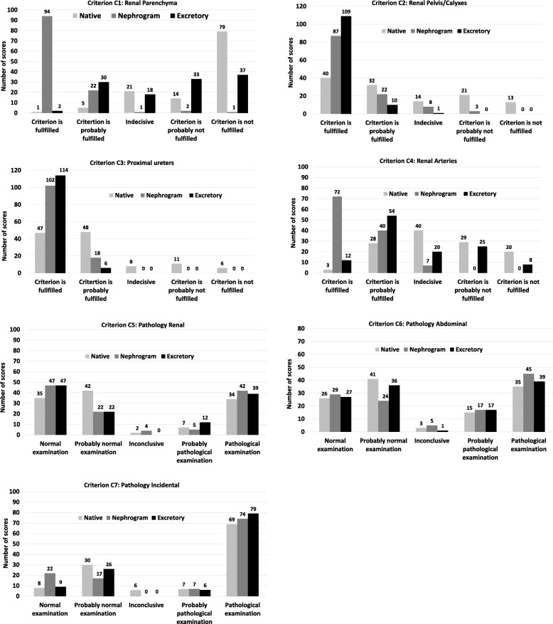
For visual reproduction of the renal parenchyma and renal arteries, the image quality was judged better for the nephrogram phase (p < 0.001), whereas renal pelvis/calyces and proximal ureters were better reproduced in the excretory phase compared to the native phase (p < 0.001). Similarly, significantly higher certainty scores were obtained in the nephrogram phase for renal parenchyma and renal arteries, but in the excretory phase for renal pelvis/calyxes and proximal ureters. Assessment of pathology in the three categories showed no statistically significant differences between the three phases. Certainty scores for assessment of pathology, however, showed a significantly higher certainty for renal pathology when comparing the native phase to nephrogram and excretory phase and a significantly higher score for nephrographic phase but only for incidental findings.
Visualisation of renal anatomy was as expected with each post-contrast phase showing favourable scores compared to the native phase. No statistically significant differences in the assessment of pathology were found between the three phases. The low-dose CT (LDCT) seems to be sufficient in differentiating between normal and pathological examinations. To reduce the radiation burden in certain patient groups, the LDCT could be considered a suitable alternative as a first line imaging method. However, radiologists should be aware of its limitations.
我们的目的是比较使用图像质量标准的原始、肾图和排泄期 CT 图像,以及在 CT 尿路造影中检测阳性病理发现,以探索是否可以降低年轻患者或结果阴性患者的辐射负担。
这是一项回顾性研究,纳入了 40 名在 192 层双源扫描仪上进行 CT 尿路造影检查的患者。根据欧洲指南,评估了四个特定的肾脏图像标准,同时评估了三个类别中的病理结果:肾脏、其他腹部和无临床意义的偶然发现。三位经验不同的放射科医生分别对每个阶段进行评估,使用分级量表。基于分级评估得出确定性评分。使用视觉分级回归(VGR)进行统计分析。显著性水平设为 p = 0.05。
在视觉再现肾脏实质和肾动脉方面,肾图期的图像质量被判断为更好(p < 0.001),而与原始期相比,排泄期肾盂/肾盏和近端输尿管的再现更好(p < 0.001)。同样,在肾图期,肾脏实质和肾动脉的确定性评分显著更高,而在排泄期,肾盂/肾盏和近端输尿管的确定性评分更高。在三个类别中评估病理学显示三个阶段之间没有统计学差异。然而,在评估病理学时,确定性评分显示,与肾图期和排泄期相比,原始期对肾脏病理学的确定性更高,并且在肾图期,只有偶然发现的评分更高。
与原始期相比,每个对比后期的肾解剖结构可视化均符合预期,显示出有利的评分。三个阶段之间的病理学评估没有统计学差异。低剂量 CT(LDCT)似乎足以区分正常和病理检查。为了降低某些患者群体的辐射负担,可以考虑将 LDCT 作为一线成像方法的合适替代方法。然而,放射科医生应该意识到其局限性。