Lerchenberger Max, Al Arabi Norah, Gallwas Julia K S, Stepp Herbert, Hallfeldt Klaus K J, Ladurner Roland
Department of Surgery, Ludwig Maximilians University Munich, Innenstadt Medical Campus, Nussbaumstrasse 20, 80336 Munich, Germany.
Department of Obstetrics and Gynecology, Ludwig Maximilians University Munich, Maistr. 11, 80337 Munich, Germany.
Int J Endocrinol. 2019 Sep 25;2019:4687951. doi: 10.1155/2019/4687951. eCollection 2019.
To investigate the feasibility of near-infrared autofluorescence (AF) and indocyanine green (ICG) fluorescence to identify parathyroid glands intraoperatively.
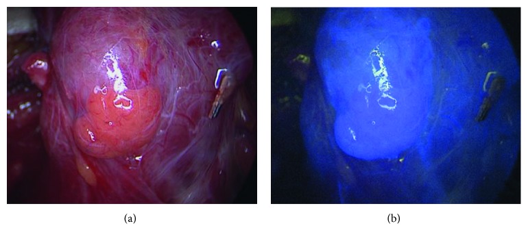
Fluorescence imaging was carried out during open parathyroid and thyroid surgery. After visual identification, parathyroid glands were exposed to near-infrared (NIR) light with a wavelength between 690 and 770 nm. The camera of the Storz® NIR/ICG endoscopic system used detects NIR light as a blue signal. Therefore, parathyroid AF was expected to be displayed in the blue color channel in contrast to the surrounding tissue. Following AF imaging, a bolus of 5 mg ICG was applied intravenously. ICG fluorescence was detected using the same NIR/ICG imaging system. Well-vascularized parathyroid glands were expected to show a strong fluorescence in contrast to surrounding lymphatic and adipose tissue.
We investigated 78 parathyroid glands from 50 patients. 64 parathyroid glands (82%) displayed AF showing the typical bluish violet color. 63 parathyroid glands (81%) showed a strong and persistent fluorescence after application of ICG. The sensitivity of identifying a parathyroid gland by AF was 82% (64 true positive and 14 false negative results), while ICG imaging showed a sensitivity of 81% (63 true positive and 15 false negative results). The Fisher exact test revealed no significant difference between both groups at < 0.05. Neither lymph nodes nor adipose tissue revealed substantial AF or ICG fluorescence.
AF and ICG fluorescence reveal a high degree of sensitivity in identifying parathyroid glands. Further, ICG imaging facilitates the assessment of parathyroid perfusion. However, in the current setting both techniques are not suitable as screening tools to identify parathyroid glands at an early stage of the operation.
探讨近红外自发荧光(AF)和吲哚菁绿(ICG)荧光在术中识别甲状旁腺的可行性。
在开放性甲状旁腺和甲状腺手术期间进行荧光成像。在视觉识别后,将甲状旁腺暴露于波长在690至770nm之间的近红外(NIR)光下。所使用的史托斯®近红外/ICG内镜系统的摄像头将近红外光检测为蓝色信号。因此,与周围组织相比,甲状旁腺AF预计会在蓝色通道中显示。AF成像后,静脉注射5mg ICG。使用相同的近红外/ICG成像系统检测ICG荧光。与周围的淋巴组织和脂肪组织相比,血运丰富的甲状旁腺预计会显示出强烈的荧光。
我们对50例患者的78个甲状旁腺进行了研究。64个甲状旁腺(82%)显示出AF,呈现典型的蓝紫色。63个甲状旁腺(81%)在注射ICG后显示出强烈且持续的荧光。通过AF识别甲状旁腺的敏感性为82%(64个真阳性和14个假阴性结果),而ICG成像的敏感性为81%(63个真阳性和15个假阴性结果)。Fisher精确检验显示两组之间在<0.05时无显著差异。淋巴结和脂肪组织均未显示出明显的AF或ICG荧光。
AF和ICG荧光在识别甲状旁腺方面显示出高度的敏感性。此外,ICG成像有助于评估甲状旁腺灌注。然而,在当前情况下,这两种技术都不适合作为在手术早期识别甲状旁腺的筛查工具。