Department of Neurosurgery, University Hospital Essen, University of Duisburg-Essen, Essen, Germany.
Institute of Neuropathology, University Hospital Essen, University of Duisburg-Essen, Essen, Germany.
BMC Neurol. 2019 Nov 23;19(1):297. doi: 10.1186/s12883-019-1529-6.
A huge spherical intracranial mass can sometimes be misdiagnosed, due to the lack of typical radiographic features. Thrombosed giant intracranial aneurysms (GIAs) are an uncommon but still a possible differential diagnosis that must be kept in mind to guarantee the best surgical approach and resection of the lesion. We describe an extremely rare case of a huge bifrontal mass mimicking a cystic echinococcosis, in which the surgery unveiled a completely thrombosed GIA of the left anterior cerebral artery (ACA).
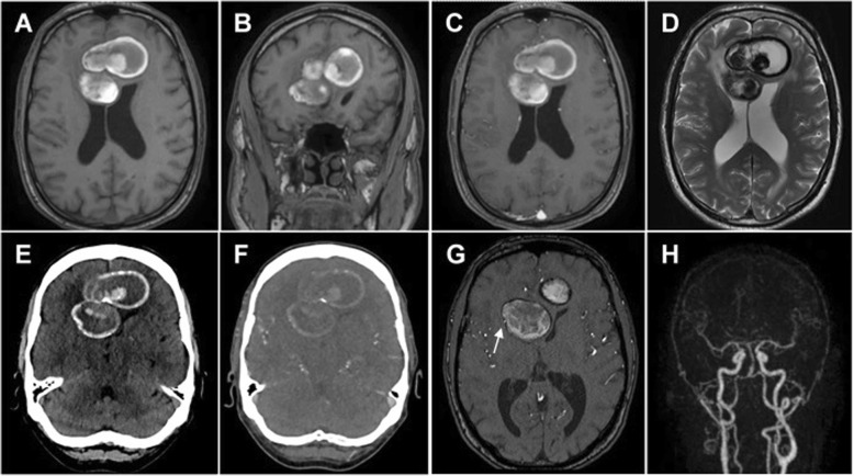
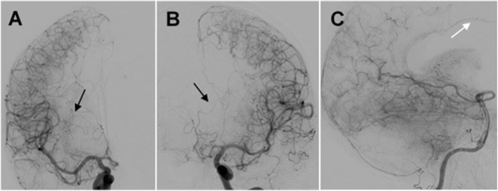
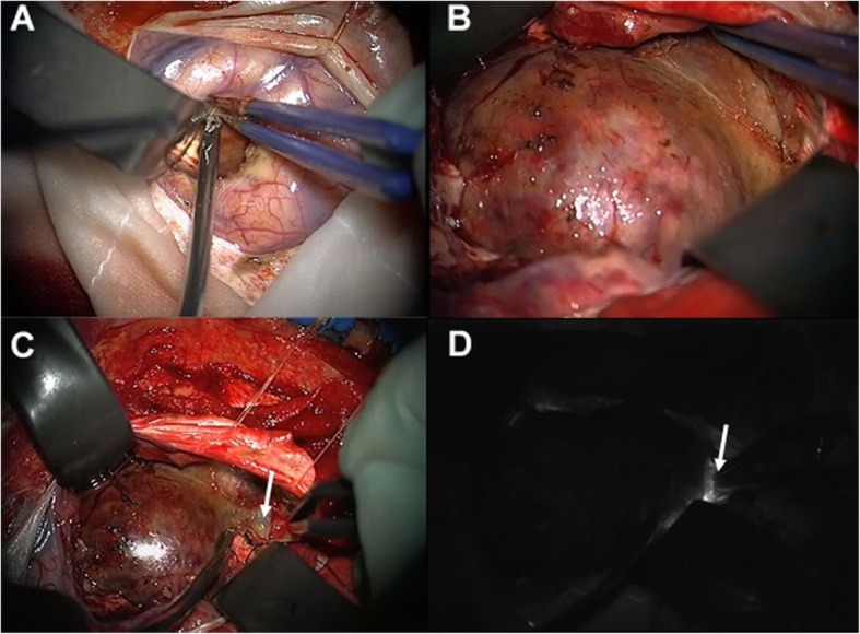
A 61-year-old patient complained about intermittent weakness of the right leg, mild holocephalic headache, beginning cognitive deficits and lethargy. Magnetic resonance imaging (MRI) showed a huge partially calcified and bilobed frontal mass with peripheral edema. Based on a time-resolved angiography with interleaved Stochastic trajectories MRI (TWIST-MRI), a vascular origin of the lesion was considered unlikely. Therefore, the surgery was performed under the suspicion of a cystic echinococcosis but revealed a bilobed GIA of the left ACA with a parent vessel thrombosis. Although only a limited left frontal craniotomy was performed, a proximal control of the parent vessel could be ensured, and the aneurysm was successfully clipped. The patient showed postoperatively no new neurological deficits.
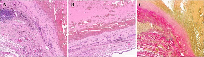
Completely thrombosed GIAs with parent vessel thrombosis are rare lesions that might be misdiagnosed if typical radiographic features are missing. Thus, in case of an intracranial spherical mass with signs of intralesional hemorrhage and mural calcifications, presence of a completely thrombosed GIA should be considered as a possible differential diagnosis.
由于缺乏典型的影像学特征,有时巨大的颅内球形肿块可能会被误诊。血栓形成的巨大颅内动脉瘤(GIAs)是一种不常见但仍需考虑的鉴别诊断,必须牢记这一点,以保证最佳的手术入路和病变切除。我们描述了一个非常罕见的病例,即一个巨大的额部肿块,类似于囊性包虫病,手术揭示了一个完全血栓形成的左侧大脑前动脉(ACA)的 GIA。
一名 61 岁患者诉右下肢间歇性无力、轻度全头痛、开始认知功能障碍和嗜睡。磁共振成像(MRI)显示一个巨大的部分钙化和双叶额部肿块,伴有周围水肿。基于时间分辨血管造影与交错随机轨迹 MRI(TWIST-MRI),病变的血管起源被认为不太可能。因此,在怀疑是囊性包虫病的情况下进行了手术,但发现左侧 ACA 的双叶 GIA 伴有母血管血栓形成。尽管仅进行了有限的左侧额骨开颅术,但可以确保近端控制母血管,并成功夹闭了动脉瘤。术后患者无新的神经功能缺损。
完全血栓形成的伴有母血管血栓形成的 GIAs 是罕见的病变,如果缺乏典型的影像学特征,可能会被误诊。因此,对于颅内球形肿块伴有瘤内出血和壁钙化的病例,如果存在完全血栓形成的 GIA,应考虑作为可能的鉴别诊断。