Research Center for Brain-inspired Intelligence and National Laboratory of Pattern Recognition, Institute of Automation, Chinese Academy of Sciences, Beijing, China; University of Chinese Academy of Sciences, Beijing, China.
Department of Anesthesia, Beijing Tongren Hospital, Capital Medical University, Beijing, China.
Neuroimage Clin. 2020;25:102188. doi: 10.1016/j.nicl.2020.102188. Epub 2020 Jan 21.
General anesthesia (GA) provides an invaluable experimental tool to understand the essential neural mechanisms underlying consciousness. Previous neuroimaging studies have shown the functional integration and segregation of brain functional networks during anesthetic-induced alteration of consciousness. However, the organization pattern of hubs in functional brain networks remains unclear. Moreover, comparisons with the well-characterized physiological unconsciousness can help us understand the neural mechanisms of anesthetic-induced unconsciousness.
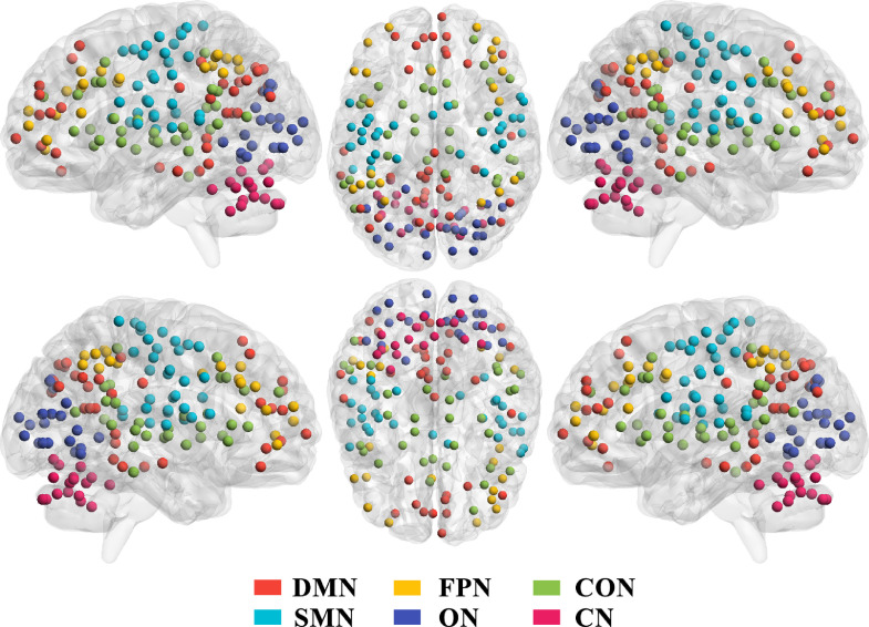
Resting-state functional magnetic resonance imaging was performed during wakefulness, mild propofol-induced sedation (m-PIS), and deep PIS (d-PIS) with clinical unconsciousness on 8 healthy volunteers and wakefulness and natural sleep on 9 age- and sex-matched healthy volunteers. Large-scale functional brain networks of each volunteer were constructed based on 160 regions of interest. Then, rich-club organizations in brain functional networks and nodal properties (nodal strength and efficiency) were assessed and analyzed among the different states and groups.
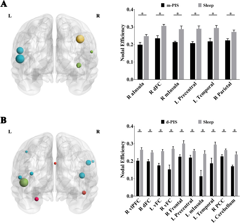
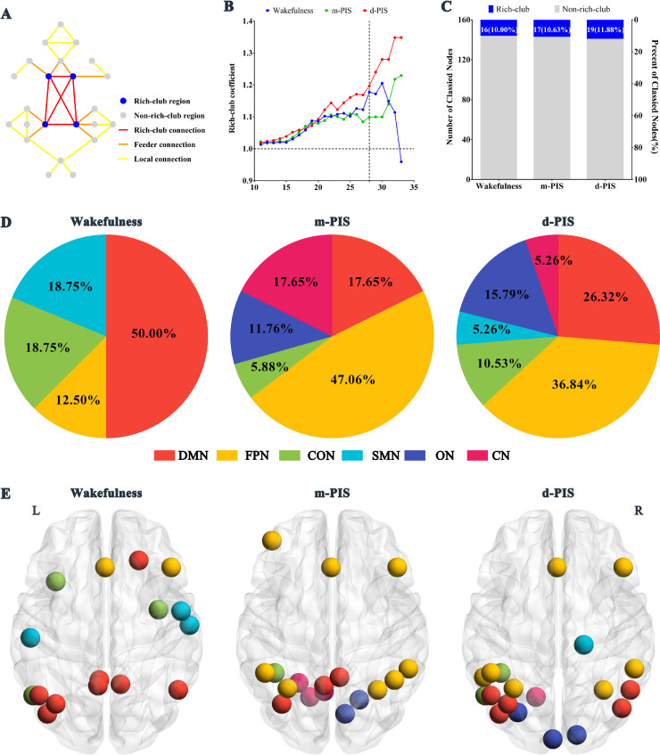
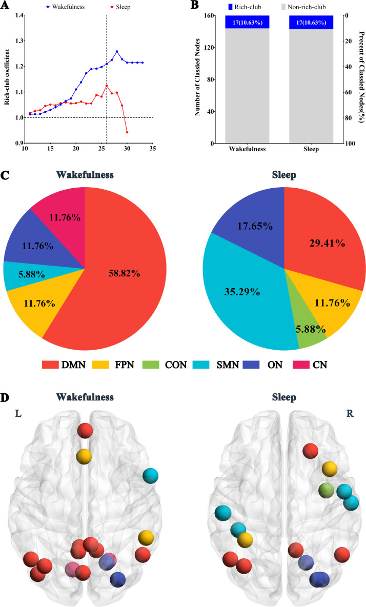
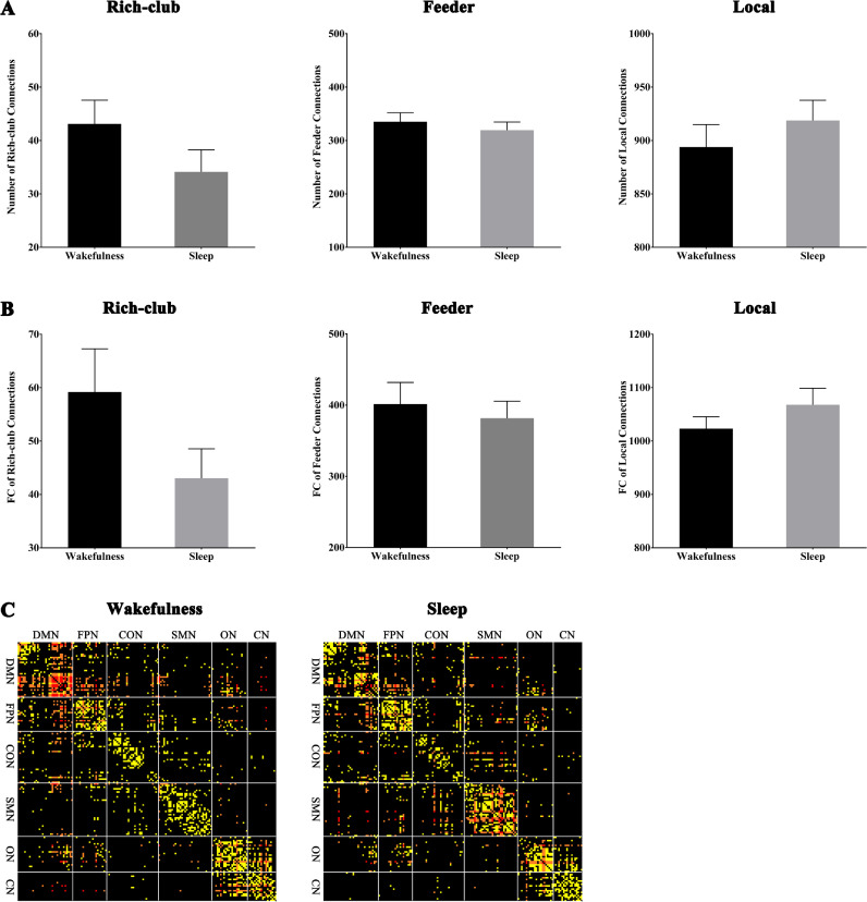
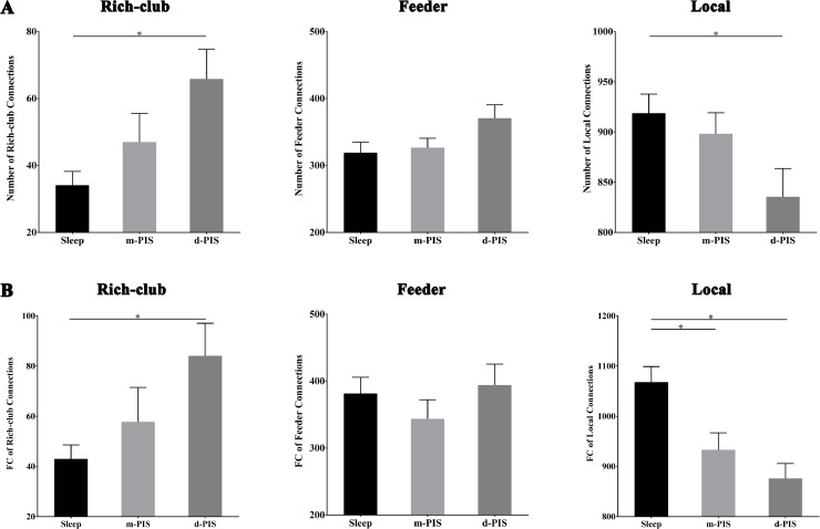
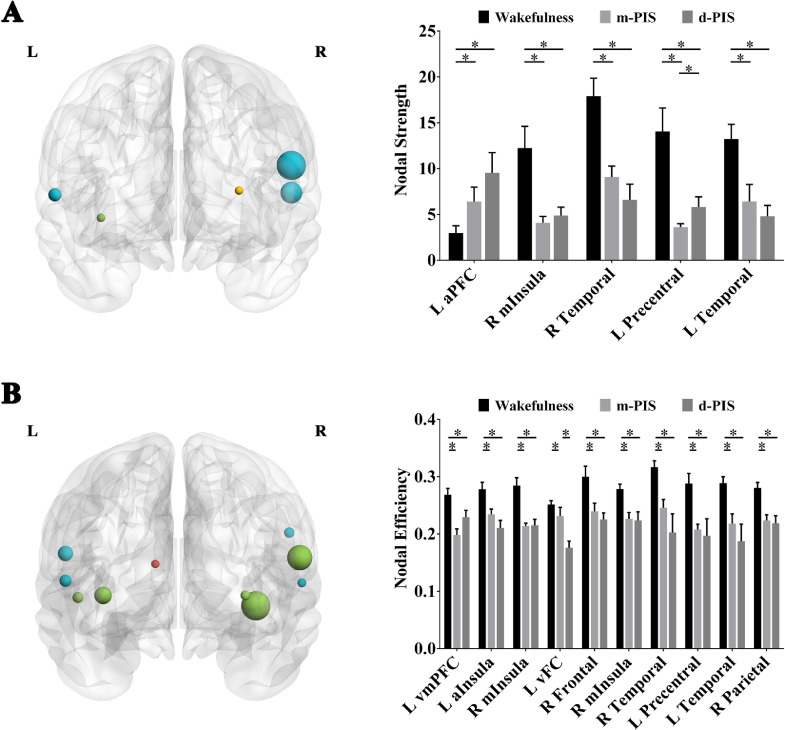
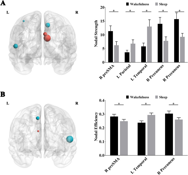
Rich-clubs in the functional brain networks were reorganized during alteration of consciousness induced by propofol. Firstly, rich-club nodes were switched from the posterior cingulate cortex (PCC), angular gyrus, and anterior and middle insula to the inferior parietal lobule (IPL), inferior parietal sulcus (IPS), and cerebellum. When sedation was deepened to unconsciousness, the rich-club nodes were switched to the occipital and angular gyrus. These results suggest that the rich-club nodes were switched among the high-order cognitive function networks (default mode network [DMN] and fronto-parietal network [FPN]), sensory networks (occipital network [ON]), and cerebellum network (CN) from consciousness (wakefulness) to propofol-induced unconsciousness. At the same time, compared with wakefulness, local connections were switched to rich-club connections during propofol-induced unconsciousness, suggesting a strengthening of the overall information commutation of networks. Nodal efficiency of the anterior and middle insula and ventral frontal cortex was significantly decreased. Additionally, from wakefulness to natural sleep, a similar pattern of rich-club reorganization with propofol-induced unconsciousness was observed: rich-club nodes were switched from the DMN (including precuneus and PCC) to the sensorimotor network (SMN, including part of the frontal and temporal gyrus). Compared with natural sleep, nodal efficiency of the insula, frontal gyrus, PCC, and cerebellum significantly decreased during propofol-induced unconsciousness.
Our study demonstrated that the rich-club reorganization in functional brain networks is characterized by switching of rich-club nodes between the high-order cognitive and sensory and motor networks during propofol-induced alteration of consciousness and natural sleep. These findings will help understand the common neurological mechanism of pharmacological and physiological unconsciousness.
全身麻醉(GA)为理解意识相关的基本神经机制提供了宝贵的实验工具。先前的神经影像学研究表明,在麻醉诱导意识改变过程中,大脑功能网络的功能整合和分离。然而,功能大脑网络枢纽的组织模式尚不清楚。此外,与特征明确的生理性无意识的比较可以帮助我们理解麻醉诱导无意识的神经机制。
在 8 名健康志愿者中,在清醒状态、轻度异丙酚诱导镇静(m-PIS)和深度 PIS(d-PIS)以及临床无意识下进行静息态功能磁共振成像,在 9 名年龄和性别匹配的健康志愿者中进行清醒和自然睡眠。基于 160 个感兴趣区构建每个志愿者的大规模功能大脑网络。然后,在不同状态和组之间评估和分析脑功能网络中的丰富俱乐部组织和节点特性(节点强度和效率)。
异丙酚诱导的意识改变过程中,功能大脑网络中的丰富俱乐部组织发生了重组。首先,丰富俱乐部节点从后扣带回皮质(PCC)、角回和前、中脑岛转移到下顶叶回(IPL)、下顶叶沟(IPS)和小脑。当镇静加深到无意识时,丰富俱乐部节点转移到枕叶和角回。这些结果表明,丰富俱乐部节点在高级认知功能网络(默认模式网络[DMN]和额顶叶网络[FPN])、感觉网络(枕叶网络[ON])和小脑网络(CN)之间切换,从意识(清醒)到异丙酚诱导的无意识。同时,与清醒相比,异丙酚诱导的无意识时,局部连接转换为丰富俱乐部连接,表明网络整体信息交换增强。前、中脑岛和腹侧额叶皮质的节点效率显著降低。此外,从清醒到自然睡眠,与异丙酚诱导的无意识时相似的丰富俱乐部重组模式被观察到:丰富俱乐部节点从 DMN(包括楔前叶和 PCC)转移到感觉运动网络(SMN,包括部分额颞叶)。与自然睡眠相比,异丙酚诱导的无意识时,脑岛、额回、PCC 和小脑的节点效率显著降低。
我们的研究表明,在异丙酚诱导的意识改变和自然睡眠过程中,功能大脑网络中的丰富俱乐部重组的特征是高级认知和感觉运动网络之间丰富俱乐部节点的切换。这些发现将有助于理解药理学和生理学无意识的共同神经机制。