Diagnostic and Interventional Radiology Department, Imam Abdulrahman Bin Faisal University, King Fahd Hospital of the University, P.O. Box: 4398, Al-Khobar City, Eastern Province, 31952, Saudi Arabia.
Diagnostic Imaging Radiology department, Royal Commission Health Services, Jubail, Saudi Arabia.
BMC Neurol. 2020 Mar 18;20(1):102. doi: 10.1186/s12883-020-01682-8.
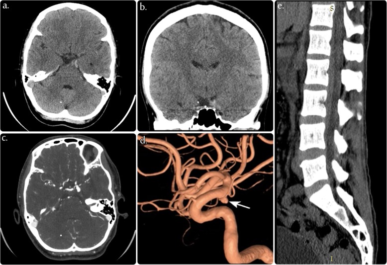
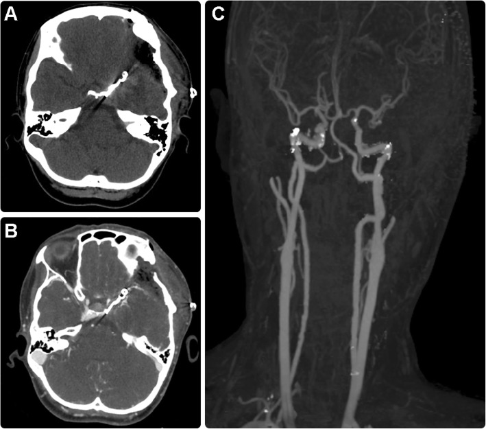
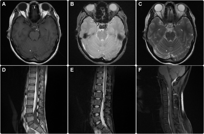
Ruptured intracranial aneurysms are often associated with subarachnoid or intraparenchymal hemorrhage. However, the prevalence of subdural hemorrhage post aneurysmal rupture is low and rarely reported in scientific studies. Here, we report an unusual case of a ruptured posterior communicating artery aneurysm resulting in an isolated subdural hematoma located in the tentorial and spinal canal without subarachnoid or intraparenchymal hemorrhage.
In this case, a 34-year-old woman with no history of trauma or coagulopathy was diagnosed with a subdural hematoma in the tentorium cerebellum tracing to the subdural space of the spinal column. Computed tomography angiography was used to identify the source of the bleeding, which revealed a ruptured left-sided posterior communicating artery saccular aneurysm. The aneurysm was clipped, and the hematoma was evacuated. The patient recovered without any neurological complications.
Our results suggest that a diagnosis of ruptured intracranial aneurysm should be considered in patients with nontraumatic subdural hematoma. Prompt diagnostic imaging and interventional diagnostic procedures are required to ensure proper management of these patients and to avoid unnecessary complications.
颅内破裂动脉瘤常伴有蛛网膜下腔或脑实质出血。然而,破裂后硬脑膜下血肿的发生率较低,在科学研究中很少报道。在此,我们报告一例罕见的后交通动脉破裂动脉瘤导致孤立性硬脑膜下血肿,位于小脑幕和椎管内,无蛛网膜下腔或脑实质出血。
本例为 34 岁女性,无外伤或凝血功能障碍史,诊断为小脑幕硬脑膜下血肿,延伸至椎管硬脑膜下间隙。计算机断层血管造影用于确定出血源,显示左侧后交通动脉囊状破裂动脉瘤。夹闭动脉瘤,清除血肿。患者无任何神经并发症恢复。
我们的结果表明,对于非外伤性硬脑膜下血肿患者,应考虑诊断为破裂颅内动脉瘤。需要及时进行诊断成像和介入诊断程序,以确保对这些患者进行适当的治疗,避免不必要的并发症。