Department of Neuroradiology, University Hospital Bonn, Venusberg-Campus 1, 53127, Bonn, Germany.
Department of Neurosurgery, University Hospital Bonn, Venusberg-Campus 1, 53127, Bonn, Germany.
Neuroradiology. 2020 Sep;62(9):1111-1122. doi: 10.1007/s00234-020-02433-9. Epub 2020 May 3.
Magnetic resonance-guided focused ultrasound (MRgFUS) systems are increasingly used to non-invasively treat tremor; consensus on imaging follow-up is poor in these patients. This study aims to elucidate how MRgFUS lesions evolve for a radiological readership with regard to clinical outcome.
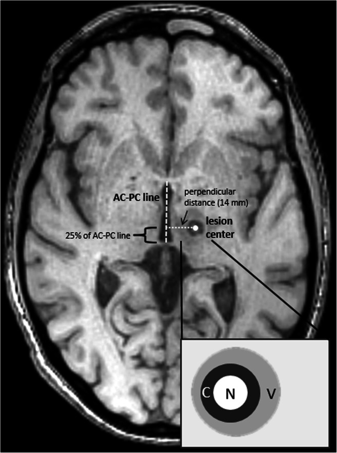
MRgFUS-induced lesions and oedema were retrospectively evaluated based on DWI, SWI, T2-weighted and T1-weighted 3-T MRI data acquired 30 min and 3, 30 and 180 days after MRgFUS (n = 9 essential tremor, n = 1 Parkinson's patients). Lesions were assessed volumetrically, visually and by ADC measurements and compared with clinical effects using non-parametric testing.
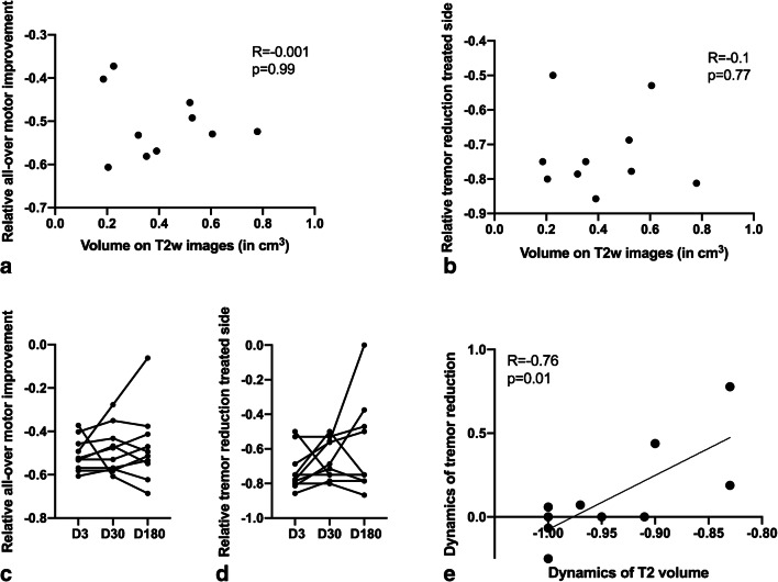
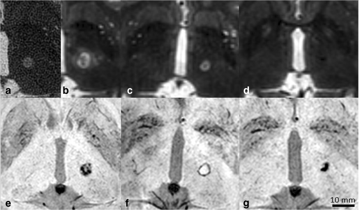
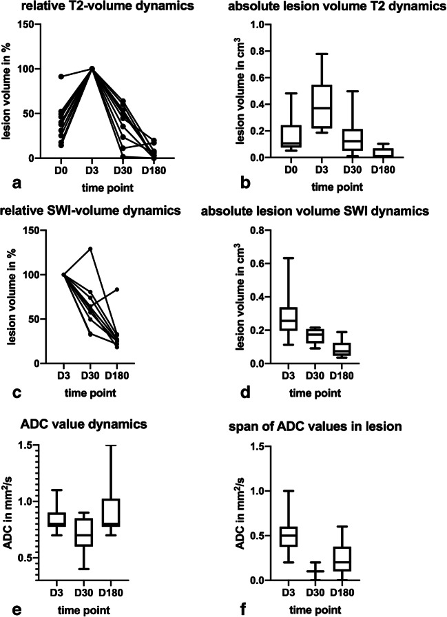
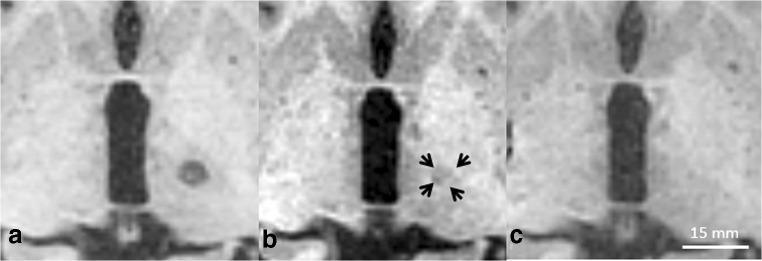
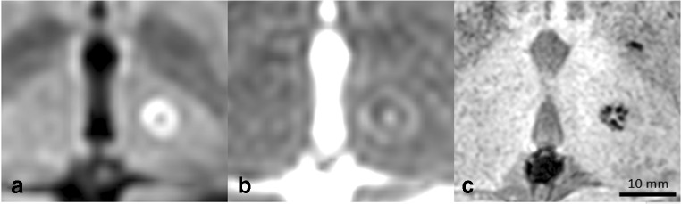
Thirty minutes after treatment, all lesions could be identified on T2-weighted images. Immediate oedema was rare (n = 1). Lesion volume as well as oedema reached a maximum on day 3 with a mean lesion size of 0.4 ± 0.2 cm and an oedema volume 3.7 ± 1.2 times the lesion volume. On day 3, a distinct diffusion-restricted rim was noted that corresponded well with SWI. Lesion shrinkage after day 3 was observed in all sequences. Lesions were no longer detectable on DWI in n = 7/10, on T2-weighted images in n = 4/10 and on T1-weighted images in n = 4/10 on day 180. No infarcts or haemorrhage were observed. There was no correlation between lesion size and initial motor skill improvement (p = 0.99). Tremor reduction dynamics correlated strongly with lesion shrinkage between days 3 and 180 (p = 0.01, R = 0.76).
In conclusion, cerebral MRgFUS lesions variably shrink over months. SWI is the sequence of choice to identify lesions after 6 months. Lesion volume is arguably associated with intermediate-term outcome.
磁共振引导聚焦超声(MRgFUS)系统越来越多地用于非侵入性地治疗震颤;但对于这些患者,影像学随访的共识很差。本研究旨在为放射科读者阐明 MRgFUS 病变如何随临床结果而演变。
根据 3T MRI 的 DWI、SWI、T2 加权和 T1 加权图像,回顾性评估了 30 分钟及治疗后 3、30 和 180 天的 MRgFUS 诱导的病变和水肿(n=9 例原发性震颤,n=1 例帕金森病患者)。通过体积评估、视觉评估和 ADC 测量评估病变,并使用非参数检验与临床效果进行比较。
治疗后 30 分钟,所有病变均能在 T2 加权图像上识别。立即出现水肿的情况很少(n=1)。病变体积和水肿在第 3 天达到最大值,平均病变大小为 0.4±0.2cm,水肿体积是病变体积的 3.7±1.2 倍。在第 3 天,观察到一个明显的弥散受限边缘,与 SWI 吻合良好。在所有序列中,第 3 天后观察到病变缩小。在第 180 天,n=7/10 的病例在 DWI 上、n=4/10 的病例在 T2 加权图像上、n=4/10 的病例在 T1 加权图像上不再检测到病变。未观察到梗死或出血。病变大小与初始运动技能改善之间无相关性(p=0.99)。震颤减轻的动力学与第 3 天至第 180 天之间的病变缩小密切相关(p=0.01,R=0.76)。
总之,脑内 MRgFUS 病变在数月内会逐渐缩小。SWI 是 6 个月后识别病变的首选序列。病变体积可能与中期结果相关。