• 文献检索
  • 文档翻译
  • 深度研究
  • 学术资讯
  • Suppr Zotero 插件Zotero 插件
  • 邀请有礼
  • 套餐&价格
  • 历史记录
应用&插件
Suppr Zotero 插件Zotero 插件浏览器插件Mac 客户端Windows 客户端微信小程序
定价
高级版会员购买积分包购买API积分包
服务
文献检索文档翻译深度研究API 文档MCP 服务
关于我们
关于 Suppr公司介绍联系我们用户协议隐私条款
关注我们

Suppr 超能文献

核心技术专利:CN118964589B侵权必究
粤ICP备2023148730 号-1Suppr @ 2026

文献检索

告别复杂PubMed语法,用中文像聊天一样搜索,搜遍4000万医学文献。AI智能推荐,让科研检索更轻松。

立即免费搜索

文件翻译

保留排版,准确专业,支持PDF/Word/PPT等文件格式,支持 12+语言互译。

免费翻译文档

深度研究

AI帮你快速写综述,25分钟生成高质量综述,智能提取关键信息,辅助科研写作。

立即免费体验

非线性光学显微镜是一种分析弹性假黄瘤皮肤改变的新工具。

Nonlinear optical microscopy is a novel tool for the analysis of cutaneous alterations in pseudoxanthoma elasticum.

机构信息

Department of Dermatology, Venereology and Dermatooncology, Semmelweis University, 41 Mária Street, Budapest, H-1085, Hungary.

Wigner RCP, Institute for Solid State Physics and Optics, Hungarian Academy of Sciences, Budapest, Hungary.

出版信息

Lasers Med Sci. 2020 Oct;35(8):1821-1830. doi: 10.1007/s10103-020-03027-w. Epub 2020 May 6.

DOI:10.1007/s10103-020-03027-w
PMID:32372237
原文链接:https://pmc.ncbi.nlm.nih.gov/articles/PMC7505829/
Abstract

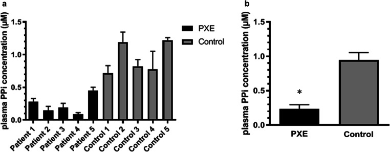
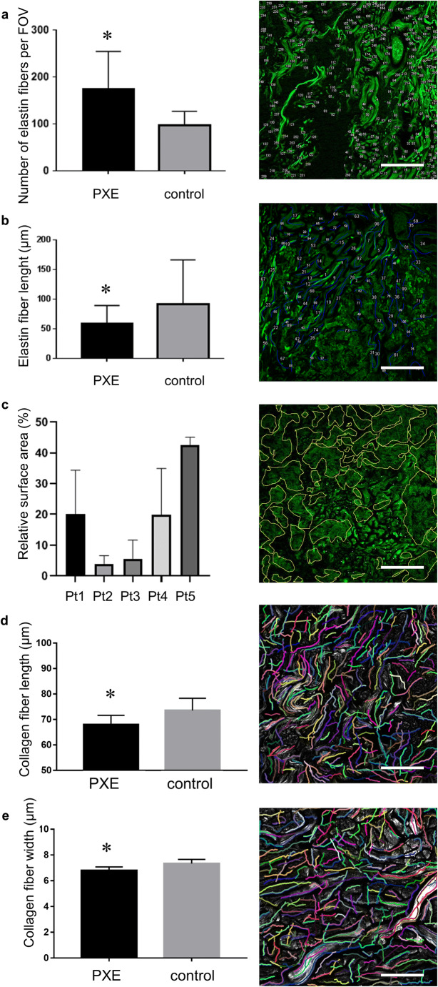
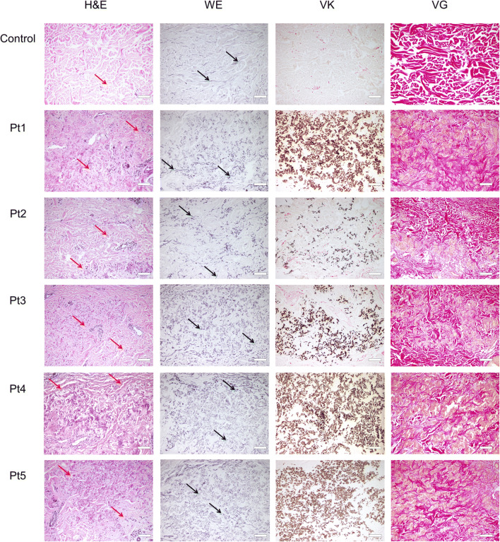
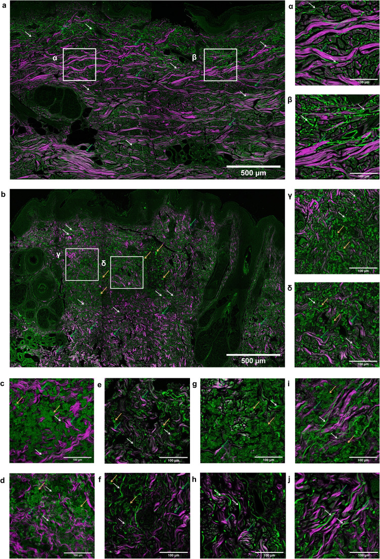
Pseudoxanthoma elasticum (PXE, OMIM 264800) is a rare autosomal recessive disorder with ectopic mineralization and fragmentation of elastin fibers. It is caused by mutations of the ABCC6 gene that leads to decreased serum levels of inorganic pyrophosphate (PPi) anti-mineralization factor. The occurrence of severe complications among PXE patients highlights the importance of early diagnosis so that prompt multidisciplinary care can be provided to patients. We aimed to examine dermal connective tissue with nonlinear optical (NLO) techniques, as collagen emits second-harmonic generation (SHG) signal, while elastin can be excited by two-photon excitation fluorescence (TPF). We performed molecular genetic analysis, ophthalmological and cardiovascular assessment, plasma PPi measurement, conventional histopathological examination, and ex vivo SHG and TPF imaging in five patients with PXE and five age- and gender-matched healthy controls. Pathological mutations including one new variant were found in the ABCC6 gene in all PXE patients and their plasma PPi level was significantly lower compared with controls. Degradation and mineralization of elastin fibers and extensive calcium deposition in the mid-dermis was visualized and quantified together with the alterations of the collagen structure in PXE. Our data suggests that NLO provides high-resolution imaging of the specific histopathological features of PXE-affected skin. In vivo NLO may be a promising tool in the assessment of PXE, promoting early diagnosis and follow-up.

摘要

弹性假黄瘤(PXE,OMIM 264800)是一种罕见的常染色体隐性遗传病,其特征为弹性纤维的异位矿化和碎裂。该病由 ABCC6 基因突变引起,导致血清中无机焦磷酸盐(PPi)抗矿化因子水平降低。PXE 患者发生严重并发症,突出了早期诊断的重要性,以便及时为患者提供多学科治疗。我们旨在使用非线性光学(NLO)技术检查皮肤结缔组织,因为胶原会发出二次谐波产生(SHG)信号,而弹性蛋白可以通过双光子激发荧光(TPF)激发。我们对 5 名 PXE 患者和 5 名年龄和性别匹配的健康对照者进行了分子遗传学分析、眼科和心血管评估、血浆 PPi 测量、常规组织病理学检查以及离体 SHG 和 TPF 成像。在所有 PXE 患者中均发现 ABCC6 基因存在病理性突变,包括一种新变异,且其血浆 PPi 水平明显低于对照组。PXE 患者的中真皮弹性纤维降解和矿化以及钙沉积广泛,胶原结构改变也可通过 NLO 成像进行可视化和定量分析。我们的数据表明 NLO 可提供 PXE 皮肤特定组织病理学特征的高分辨率成像。体内 NLO 可能成为 PXE 评估的一种有前途的工具,有助于早期诊断和随访。

https://cdn.ncbi.nlm.nih.gov/pmc/blobs/94d2/7505829/b94d204e05ea/10103_2020_3027_Fig4_HTML.jpg
https://cdn.ncbi.nlm.nih.gov/pmc/blobs/94d2/7505829/c18dfba48114/10103_2020_3027_Fig1_HTML.jpg
https://cdn.ncbi.nlm.nih.gov/pmc/blobs/94d2/7505829/099c2b8addab/10103_2020_3027_Fig2_HTML.jpg
https://cdn.ncbi.nlm.nih.gov/pmc/blobs/94d2/7505829/7c81c4911dc5/10103_2020_3027_Fig3_HTML.jpg
https://cdn.ncbi.nlm.nih.gov/pmc/blobs/94d2/7505829/b94d204e05ea/10103_2020_3027_Fig4_HTML.jpg

相似文献

1
Nonlinear optical microscopy is a novel tool for the analysis of cutaneous alterations in pseudoxanthoma elasticum.非线性光学显微镜是一种分析弹性假黄瘤皮肤改变的新工具。
Lasers Med Sci. 2020 Oct;35(8):1821-1830. doi: 10.1007/s10103-020-03027-w. Epub 2020 May 6.
2
Correlation of systemic involvement and presence of pathological skin calcification assessed by ex vivo nonlinear microscopy in Pseudoxanthoma elasticum.体外非线性显微镜评估弹性假黄瘤系统受累与病理性皮肤钙化的相关性。
Arch Dermatol Res. 2023 Sep;315(7):1897-1908. doi: 10.1007/s00403-023-02557-x. Epub 2023 Feb 27.
3
Immunochemical identification of abnormal constituents in the dermis of pseudoxanthoma elasticum patients.弹性假黄瘤患者真皮中异常成分的免疫化学鉴定
Eur J Histochem. 1994;38(2):111-23.
4
Pseudoxanthoma elasticum.弹性假黄瘤
Orphanet J Rare Dis. 2017 May 10;12(1):85. doi: 10.1186/s13023-017-0639-8.
5
Atorvastatin counteracts aberrant soft tissue mineralization in a mouse model of pseudoxanthoma elasticum (Abcc6⁻/⁻).阿托伐他汀可逆转弹性假黄瘤(Abcc6⁻/⁻)小鼠模型中的软组织矿化异常。
J Mol Med (Berl). 2013 Oct;91(10):1177-84. doi: 10.1007/s00109-013-1066-5. Epub 2013 Jun 27.
6
Serum factors from pseudoxanthoma elasticum patients alter elastic fiber formation in vitro.弹性假黄瘤患者的血清因子在体外会改变弹性纤维的形成。
J Invest Dermatol. 2006 Jul;126(7):1497-505. doi: 10.1038/sj.jid.5700201. Epub 2006 Mar 16.
7
Ex vivo nonlinear microscopy imaging of Ehlers-Danlos syndrome-affected skin.体外非线性显微镜成像技术在埃勒斯-当洛斯综合征皮肤病变中的应用。
Arch Dermatol Res. 2018 Jul;310(5):463-473. doi: 10.1007/s00403-018-1835-7. Epub 2018 May 3.
8
The mineralization of elastic fibers and alterations of extracellular matrix in pseudoxanthoma elasticum. Ultrastructure, immunocytochemistry, and X-ray analysis.弹性假黄瘤中弹性纤维的矿化及细胞外基质的改变。超微结构、免疫细胞化学及X射线分析。
Arch Dermatol. 1989 Jan;125(1):70-6.
9
Pyrophosphates as a major inhibitor of matrix calcification in Pseudoxanthoma elasticum.焦磷酸盐作为弹性假黄瘤中基质钙化的主要抑制剂。
J Dermatol Sci. 2014 Aug;75(2):109-20. doi: 10.1016/j.jdermsci.2014.04.015. Epub 2014 May 17.
10
Fibroblasts from patients affected by Pseudoxanthoma elasticum exhibit an altered PPi metabolism and are more responsive to pro-calcifying stimuli.患有弹性假黄瘤的患者的成纤维细胞表现出焦磷酸(PPi)代谢改变,并且对促钙化刺激反应更强。
J Dermatol Sci. 2014 Apr;74(1):72-80. doi: 10.1016/j.jdermsci.2013.12.008. Epub 2014 Jan 2.

引用本文的文献

1
A Cross-Sectional Study of the Dermatological Manifestations of Patients with Fabry Disease and the Assessment of Angiokeratomas with Multimodal Imaging.法布里病患者皮肤表现的横断面研究及血管角质瘤的多模态成像评估
Diagnostics (Basel). 2023 Jul 14;13(14):2368. doi: 10.3390/diagnostics13142368.
2
[Dermoscopy of genodermatoses].[遗传性皮肤病的皮肤镜检查]
Dermatologie (Heidelb). 2023 Apr;74(4):256-261. doi: 10.1007/s00105-023-05124-7. Epub 2023 Mar 7.
3
Correlation of systemic involvement and presence of pathological skin calcification assessed by ex vivo nonlinear microscopy in Pseudoxanthoma elasticum.

本文引用的文献

1
Nonlinear microscopy of common histological stains reveals third harmonic generation harmonophores.常见组织学染色的非线性显微镜显示三谐波产生的类晶。
Analyst. 2019 May 13;144(10):3239-3249. doi: 10.1039/c9an00267g.
2
Endogenous Calcification Inhibitors in the Prevention of Vascular Calcification: A Consensus Statement From the COST Action EuroSoftCalcNet.内源性钙化抑制剂在预防血管钙化中的作用:欧洲软钙化网络(COST行动)共识声明
Front Cardiovasc Med. 2019 Jan 18;5:196. doi: 10.3389/fcvm.2018.00196. eCollection 2018.
3
Multiphoton Imaging of Collagen, Elastin, and Calcification in Intact Soft-Tissue Samples.
体外非线性显微镜评估弹性假黄瘤系统受累与病理性皮肤钙化的相关性。
Arch Dermatol Res. 2023 Sep;315(7):1897-1908. doi: 10.1007/s00403-023-02557-x. Epub 2023 Feb 27.
4
Arterial dissections: Common features and new perspectives.动脉夹层:共同特征与新观点
Front Cardiovasc Med. 2022 Dec 6;9:1055862. doi: 10.3389/fcvm.2022.1055862. eCollection 2022.
5
A new enzymatic assay to quantify inorganic pyrophosphate in plasma.一种用于定量检测血浆中无机焦磷酸的新酶分析法。
Anal Bioanal Chem. 2023 Jan;415(3):481-492. doi: 10.1007/s00216-022-04430-8. Epub 2022 Nov 19.
6
Serum Calcification Propensity T50 Associates with Disease Severity in Patients with Pseudoxanthoma Elasticum.血清钙化倾向T50与弹性假黄瘤患者的疾病严重程度相关。
J Clin Med. 2022 Jun 28;11(13):3727. doi: 10.3390/jcm11133727.
7
Polarized photoacoustic microscopy based on high-order harmonics for anisotropy detection.基于高阶谐波的偏振光声显微镜用于各向异性检测。
Ann Transl Med. 2022 Mar;10(6):293. doi: 10.21037/atm-22-794.
8
Multiphoton microscopy providing pathological-level quantification of myocardial fibrosis in transplanted human heart.多光子显微镜对移植人心肌纤维化进行病理水平的定量分析。
Lasers Med Sci. 2022 Sep;37(7):2889-2898. doi: 10.1007/s10103-022-03557-5. Epub 2022 Apr 8.
9
Low concentration staining for high chemical contrast, nonlinear microscope mosaic imaging of skin alterations in pseudoxanthoma elasticum.用于高化学对比度的低浓度染色,弹性假黄瘤皮肤改变的非线性显微镜镶嵌成像。
Biomed Opt Express. 2021 Dec 8;13(1):252-261. doi: 10.1364/BOE.443507. eCollection 2022 Jan 1.
10
Artificial Intelligence-Based Approaches to Reflectance Confocal Microscopy Image Analysis in Dermatology.基于人工智能的皮肤科反射共聚焦显微镜图像分析方法
J Clin Med. 2022 Jan 14;11(2):429. doi: 10.3390/jcm11020429.
完整软组织样本中胶原蛋白、弹性蛋白和钙化的多光子成像
Curr Protoc Cytom. 2019 Jan;87(1):e51. doi: 10.1002/cpcy.51. Epub 2018 Oct 31.
4
The extent of pseudoxanthoma elasticum skin changes is related to cardiovascular complications and visual loss: a cross-sectional study.弹性假黄瘤皮肤改变的程度与心血管并发症及视力丧失相关:一项横断面研究。
Br J Dermatol. 2019 Jan;180(1):207-208. doi: 10.1111/bjd.17094. Epub 2018 Oct 14.
5
Quantitative Analysis on Ex Vivo Nonlinear Microscopy Images of Basal Cell Carcinoma Samples in Comparison to Healthy Skin.体外对比健康皮肤的基底细胞癌样本的非线性显微镜图像的定量分析
Pathol Oncol Res. 2019 Jul;25(3):1015-1021. doi: 10.1007/s12253-018-0445-1. Epub 2018 Jul 6.
6
The prevalence of pseudoxanthoma elasticum: Revised estimations based on genotyping in a high vascular risk cohort.弹性假黄瘤的患病率:基于高血管风险队列基因分型的修订估计值。
Eur J Med Genet. 2019 Feb;62(2):90-92. doi: 10.1016/j.ejmg.2018.05.020. Epub 2018 May 22.
7
Ex vivo nonlinear microscopy imaging of Ehlers-Danlos syndrome-affected skin.体外非线性显微镜成像技术在埃勒斯-当洛斯综合征皮肤病变中的应用。
Arch Dermatol Res. 2018 Jul;310(5):463-473. doi: 10.1007/s00403-018-1835-7. Epub 2018 May 3.
8
Alteration of Extracellular Nucleotide Metabolism in Pseudoxanthoma Elasticum.弹性假黄瘤中外切核苷酸代谢的改变。
J Invest Dermatol. 2018 Aug;138(8):1862-1870. doi: 10.1016/j.jid.2018.02.023. Epub 2018 Mar 6.
9
Non-destructive two-photon excited fluorescence imaging identifies early nodules in calcific aortic-valve disease.非破坏性双光子激发荧光成像可识别钙化性主动脉瓣疾病中的早期结节。
Nat Biomed Eng. 2017 Nov;1(11):914-924. doi: 10.1038/s41551-017-0152-3. Epub 2017 Nov 6.
10
Stain-free Histopathology of Basal Cell Carcinoma by Dual Vibration Resonance Frequency CARS Microscopy.基于双振动共振频率相干反斯托克斯拉曼散射显微镜的基底细胞癌无标记组织病理学研究
Pathol Oncol Res. 2018 Oct;24(4):927-930. doi: 10.1007/s12253-017-0356-6. Epub 2017 Nov 4.