Department of Neurology, Odense University Hospital, Odense, Denmark.
Neurology Research Unit, Department of Clinical Research, University of Southern Denmark, Odense, Denmark.
Clin Exp Immunol. 2020 Sep;201(3):328-340. doi: 10.1111/cei.13473. Epub 2020 Jul 6.
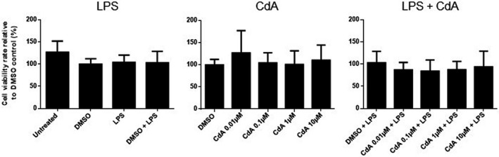
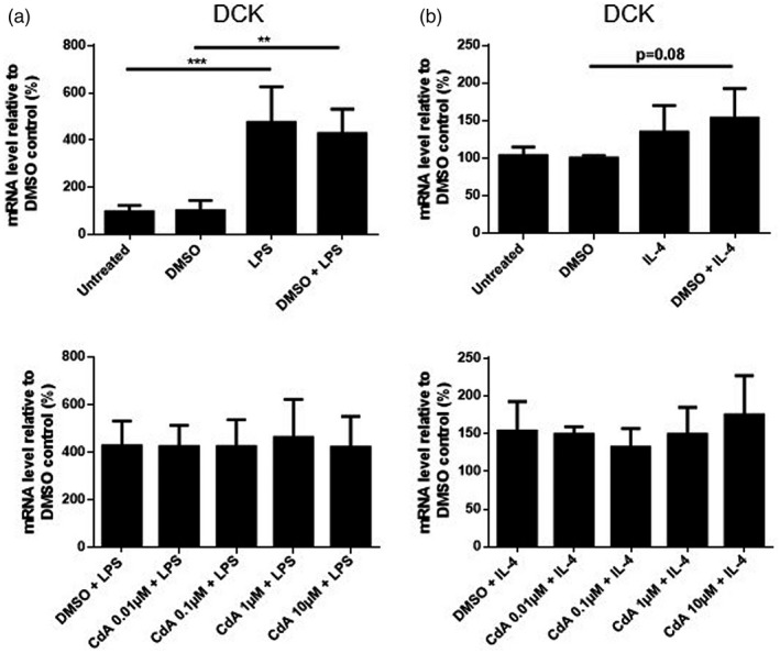
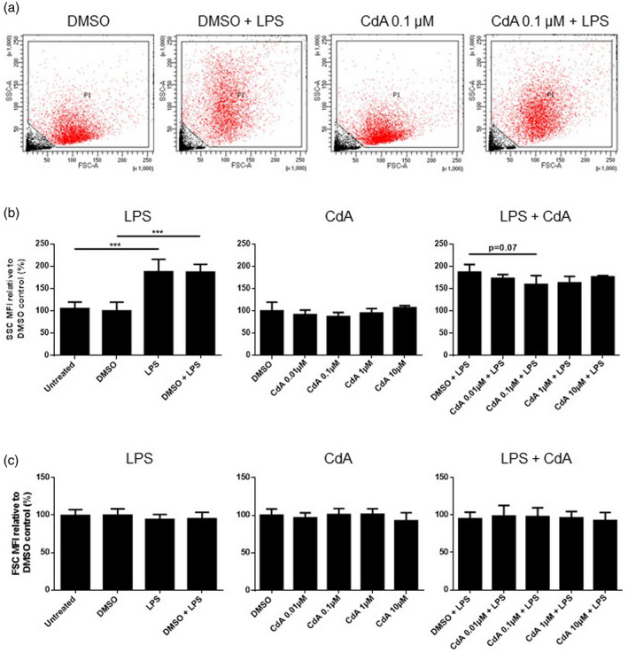
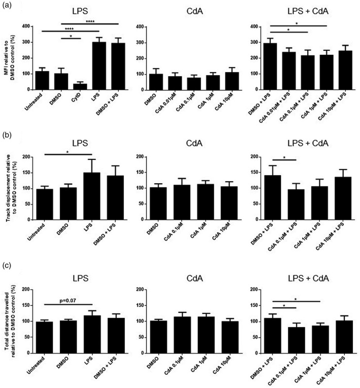
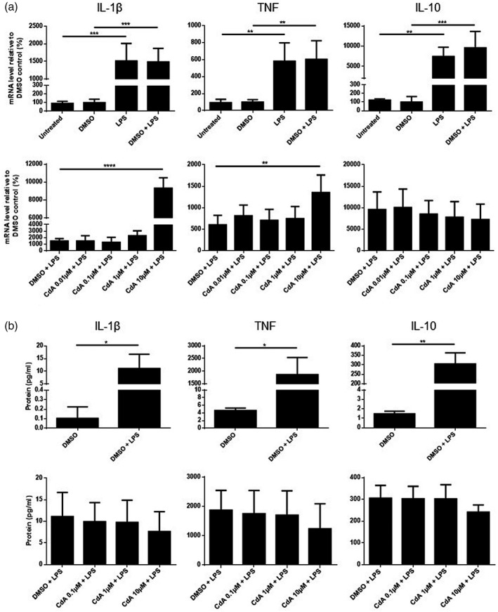
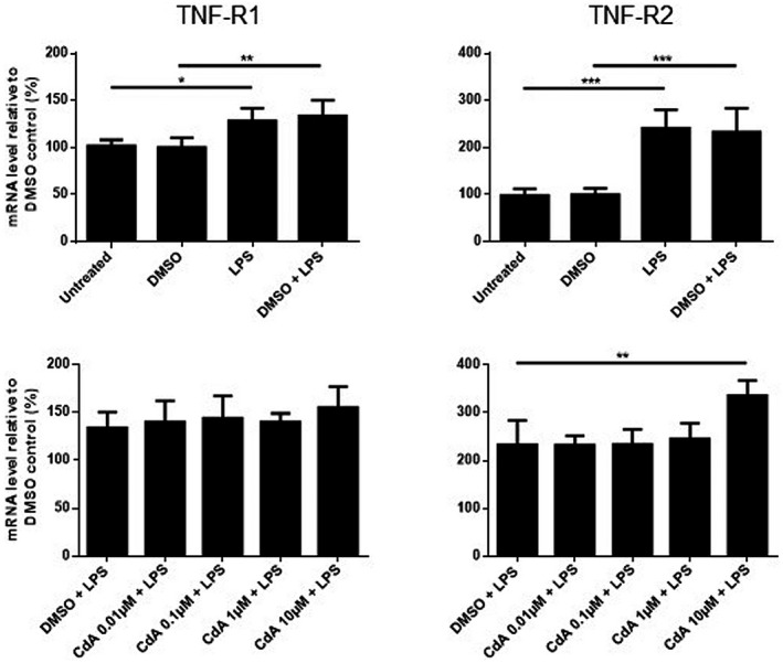
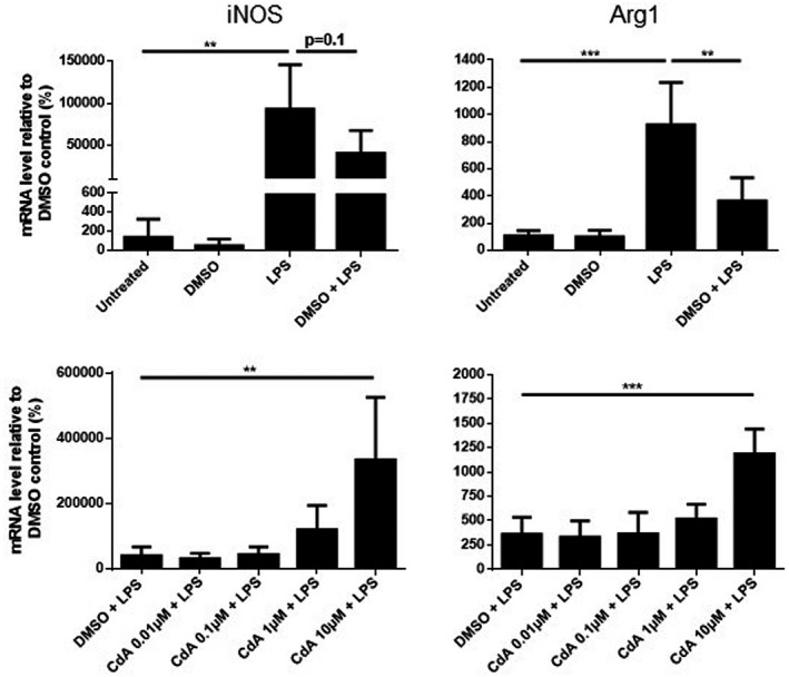
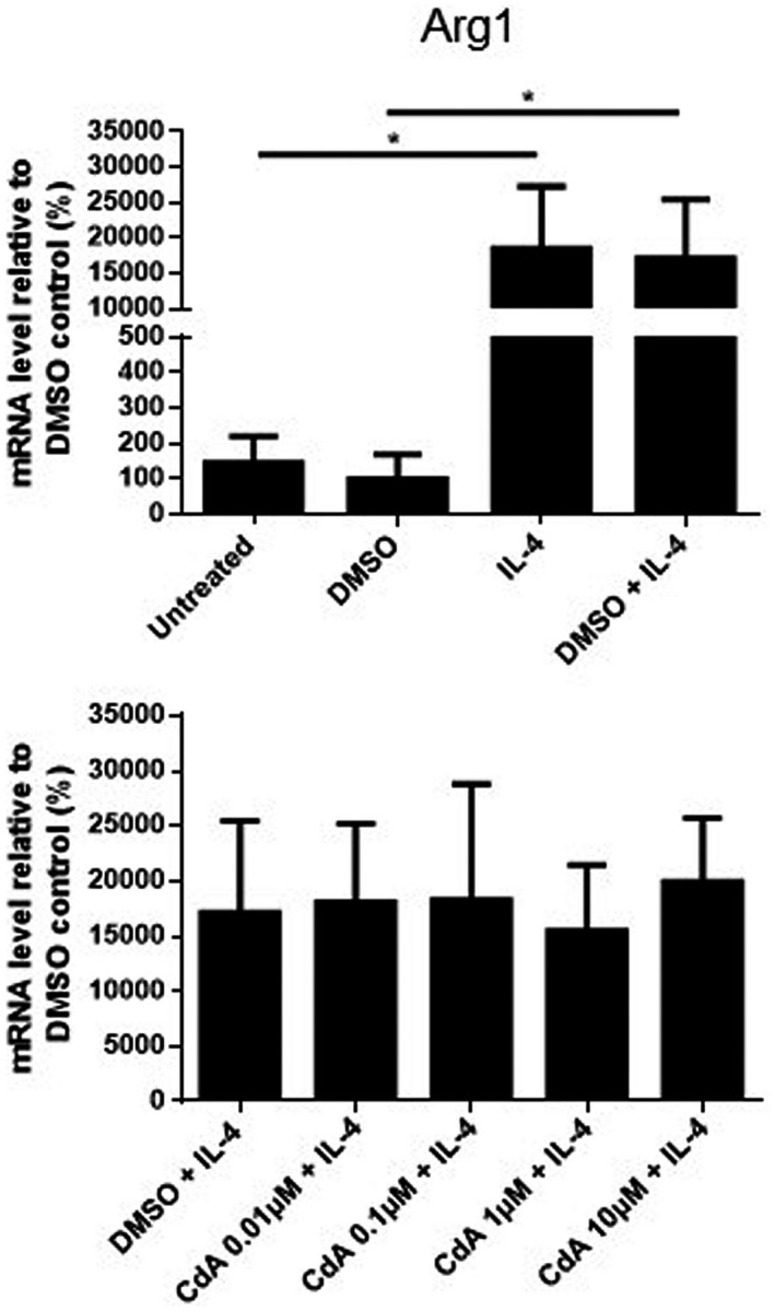
Cladribine (CdA), an oral prodrug approved for the treatment of relapsing multiple sclerosis, selectively depletes lymphocytes. CdA passes the blood-brain barrier, suggesting a potential effect on central nervous system (CNS) resident cells. We examined if CdA modifies the phenotype and function of naive and activated primary mouse microglia, when applied in the concentrations 0·1-1 μM that putatively overlap human cerebrospinal fluid (CSF) concentrations. Primary microglia cultures without stimulation or in the presence of proinflammatory lipopolysaccharide (LPS) or anti-inflammatory interleukin (IL)-4 were treated with different concentrations of CdA for 24 h. Viability was assessed by MTT [3-(4,5-dimethylthiazol-2-yl)-2,5-diphenyltetrazolium bromide] assay. Phagocytotic ability and morphology were examined by flow cytometry and random migration using IncuCyte Zoom and TrackMate. Change in gene expression was examined by quantitative polymerase chain reaction (qPCR) and protein secretion by Meso Scale Discovery. We found that LPS and IL-4 up-regulated deoxycytidine kinase (DCK) expression. Only activated microglia were affected by CdA, and this was unrelated to viability. CdA 0·1-1 μM significantly reduced granularity, phagocytotic ability and random migration of activated microglia. CdA 10 μM increased the IL-4-induced gene expression of arginase 1 (Arg1) and LPS-induced expression of IL-1β, tumor necrosis factor (TNF), inducible nitric oxide synthase (iNOS) and Arg1, but protein secretion remained unaffected. CdA 10 μM potentiated the increased expression of anti-inflammatory TNF receptor 2 (TNF-R2) but not TNF-R1 induced by LPS. This suggests that microglia acquire a less activated phenotype when treated with 0·1-1 μM CdA that putatively overlaps human CSF concentrations. This may be related to the up-regulated gene expression of DCK upon activation, and suggests a potential alternative mechanism of CdA with direct effect on CNS resident cells.
克拉屈滨(CdA)是一种口服前体药物,用于治疗复发性多发性硬化症,选择性地消耗淋巴细胞。CdA 能够穿过血脑屏障,这表明它可能对中枢神经系统(CNS)固有细胞产生影响。我们研究了当 CdA 以 0.1-1 μM 的浓度应用于无刺激或存在促炎脂多糖(LPS)或抗炎白细胞介素(IL)-4 的情况下,是否会改变幼稚和激活的原代小鼠小胶质细胞的表型和功能,这些浓度推测与人类脑脊液(CSF)浓度重叠。未经刺激或存在促炎 LPS 或抗炎 IL-4 的原代小胶质细胞培养物用不同浓度的 CdA 处理 24 小时。通过 MTT [3-(4,5-二甲基噻唑-2-基)-2,5-二苯基四唑溴化物]测定法评估细胞活力。通过流式细胞术和 IncuCyte Zoom 和 TrackMate 检测吞噬能力和形态。通过定量聚合酶链反应(qPCR)和 Meso Scale Discovery 检测蛋白质分泌来检查基因表达的变化。我们发现 LPS 和 IL-4 上调脱氧胞苷激酶(DCK)的表达。只有激活的小胶质细胞受到 CdA 的影响,而这与活力无关。0.1-1 μM 的 CdA 显著降低了激活的小胶质细胞的颗粒度、吞噬能力和随机迁移。10 μM 的 CdA 增加了 IL-4 诱导的精氨酸酶 1(Arg1)基因表达和 LPS 诱导的白细胞介素 1β(IL-1β)、肿瘤坏死因子(TNF)、诱导型一氧化氮合酶(iNOS)和 Arg1 的表达,但蛋白质分泌不受影响。10 μM 的 CdA 增强了 LPS 诱导的抗炎 TNF 受体 2(TNF-R2)的表达,但没有增强 TNF-R1 的表达。这表明,当用 0.1-1 μM 的 CdA 处理时,小胶质细胞获得了一种较少激活的表型,这种表型推测与人类 CSF 浓度重叠。这可能与激活时 DCK 的基因表达上调有关,并提示 CdA 可能具有一种直接作用于中枢神经系统固有细胞的潜在替代机制。