Montanaro Domenico, Vavla M, Frijia F, Aghakhanyan G, Baratto A, Coi A, Stefan C, Girardi G, Paparella G, De Cori S, Totaro P, Lombardo F, Piccoli G, Martinuzzi Andrea
U.O.C. Risonanza Magnetica Specialistica e Neuroradiologia, Fondazione CNR/Regione Toscana G. Monasterio, Pisa, Italy.
Severe Developmental Disabilities Unit, Scientific Institute, IRCCS Eugenio Medea, Conegliano, Italy.
Front Neurosci. 2020 Jun 4;14:325. doi: 10.3389/fnins.2020.00325. eCollection 2020.
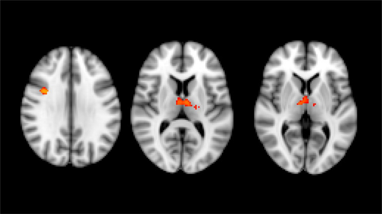
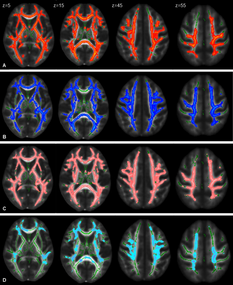
Hereditary spastic paraplegias (HSP) are a group of genetically and clinically heterogeneous neurologic disorders. Hereby we describe a relatively large group of patients (pts) affected by HSP studied at baseline (31 pts) and at follow-up (mean period 28.9 ± 8.4 months; 23 pts) with multimodal advanced MRI: high-resolution T1 images for voxel-based morphometry (VBM) analysis, magnetic resonance spectroscopy (MRS), and diffusion tensor imaging (DTI). An age-matched healthy control (HC) group underwent the same neuroimaging protocol in a time schedule matched with the HSP patients. At baseline, VBM showed gray matter (GM) reduction in HSP in the right pre-frontal cortex and bilaterally in the thalami. MRS at baseline depicted in HSP patients compared to the HC group reduction of NAA/Cr ratio in the right pre-frontal region, increase of Cho/Cr ratio in the right pre-central regions, and increase of mI/Cr ratio on the left pre-central area. At cross-sectional follow-up analysis and longitudinal evaluation, no VBM and MRS statistically significant results were obtained. Tract-based spatial statistics (TBSS) analysis showed widespread DTI brain white matter (WM) alterations in patients compared to HC at baseline, which are characterized by reduction of fractional anisotropy (FA) and increase of mean diffusivity (MD), axial diffusivity (AD), and radial diffusivity, as confirmed on cross-analysis of the follow-up dataset. A longitudinal analysis with TBSS in HSP patients did not show significant variations, while upon applying region-based analysis we found increased FA and decreased MD and AD in specific brain WM fiber complex during follow-up. The changes were not correlated with the clinical presentation (pure complicated HSP), motor function, and motility indexes or history of specific treatments (botulinum toxin). In conclusion, the cross-sectional analysis of the multiparametric MRI data in our HSP patients confirmed the non-prominent involvement of the cortex in the primary motor regions but rather of other more associative areas. On the contrary, DTI demonstrated a widespread involvement of the brain WM, including the primary motor regions, which was confirmed at follow-up. The longitudinal analysis revealed an apparent inversion of tendency when considering the expected evolution of a neurodegenerative process: we detected an increase of FA and a decrease of MD and AD. These time-related modifications may suggest a repair attempt by the residual central WM fibers, which requires confirmation with a larger group of patients and with a longer time interval.
遗传性痉挛性截瘫(HSP)是一组在遗传和临床方面具有异质性的神经疾病。在此,我们描述了一组相对较大的受HSP影响的患者,在基线时(31例)以及随访时(平均时间28.9±8.4个月;23例)进行了多模态先进磁共振成像(MRI)检查:用于基于体素的形态学测量(VBM)分析的高分辨率T1图像、磁共振波谱(MRS)以及扩散张量成像(DTI)。一个年龄匹配的健康对照组(HC)按照与HSP患者相匹配的时间安排接受了相同的神经影像学检查方案。在基线时,VBM显示HSP患者右侧前额叶皮质以及双侧丘脑的灰质(GM)减少。与HC组相比,HSP患者基线时的MRS显示右侧前额叶区域NAA/Cr比值降低,右侧中央前区域Cho/Cr比值升高,左侧中央前区域mI/Cr比值升高。在横断面随访分析和纵向评估中,未获得VBM和MRS具有统计学意义的结果。基于纤维束的空间统计学(TBSS)分析显示,与基线时的HC组相比,患者的DTI脑白质(WM)存在广泛改变,其特征为分数各向异性(FA)降低以及平均扩散率(MD)、轴向扩散率(AD)和径向扩散率升高,这在随访数据集的交叉分析中得到了证实。HSP患者的TBSS纵向分析未显示出显著变化,而在应用基于区域的分析时,我们发现在随访期间特定脑WM纤维复合体中的FA升高,MD和AD降低。这些变化与临床表现(单纯型/复杂型HSP)、运动功能、活动指数或特定治疗史(肉毒杆菌毒素)无关。总之,我们对HSP患者多参数MRI数据的横断面分析证实,初级运动区域的皮质受累并不突出,而其他更多的联合区域受累更为明显。相反,DTI显示脑WM广泛受累,包括初级运动区域,这在随访中得到了证实。纵向分析显示,在考虑神经退行性过程的预期演变时出现了明显的趋势反转:我们检测到FA升高,MD和AD降低。这些与时间相关的改变可能提示残留的中枢WM纤维有修复尝试,这需要在更大规模的患者群体以及更长的时间间隔内进行证实。