• 文献检索
  • 文档翻译
  • 深度研究
  • 学术资讯
  • Suppr Zotero 插件Zotero 插件
  • 邀请有礼
  • 套餐&价格
  • 历史记录
应用&插件
Suppr Zotero 插件Zotero 插件浏览器插件Mac 客户端Windows 客户端微信小程序
定价
高级版会员购买积分包购买API积分包
服务
文献检索文档翻译深度研究API 文档MCP 服务
关于我们
关于 Suppr公司介绍联系我们用户协议隐私条款
关注我们

Suppr 超能文献

核心技术专利:CN118964589B侵权必究
粤ICP备2023148730 号-1Suppr @ 2026

文献检索

告别复杂PubMed语法,用中文像聊天一样搜索,搜遍4000万医学文献。AI智能推荐,让科研检索更轻松。

立即免费搜索

文件翻译

保留排版,准确专业,支持PDF/Word/PPT等文件格式,支持 12+语言互译。

免费翻译文档

深度研究

AI帮你快速写综述,25分钟生成高质量综述,智能提取关键信息,辅助科研写作。

立即免费体验

拇外翻畸形中的跖骨内翻:综述。

Metatarsal Pronation in Hallux Valgus Deformity: A Review.

机构信息

Departamento de Traumatologia, Clinica Alemana-Universidad del Desarrollo, Santiago, Chile.

出版信息

J Am Acad Orthop Surg Glob Res Rev. 2020 Jun 15;4(6). doi: 10.5435/JAAOSGlobal-D-20-00091. eCollection 2020 Jun.

DOI:10.5435/JAAOSGlobal-D-20-00091
PMID:32656482
原文链接:https://pmc.ncbi.nlm.nih.gov/articles/PMC7322783/
Abstract

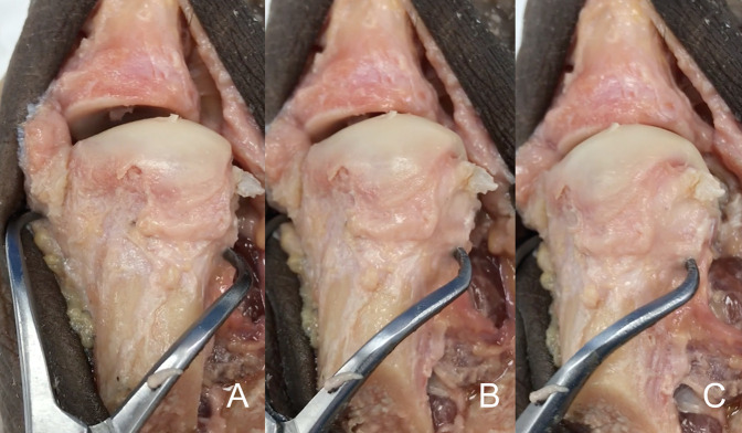
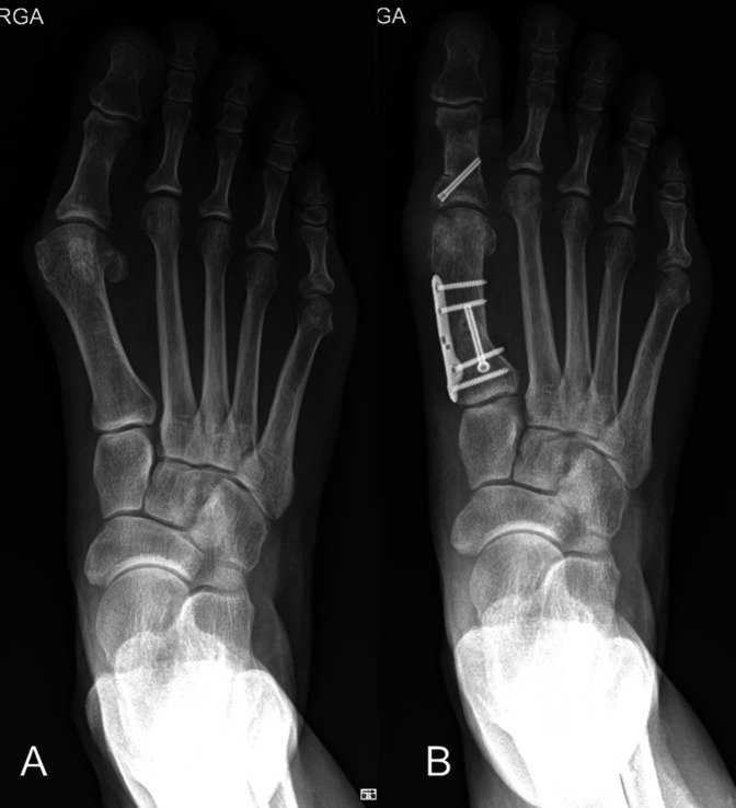
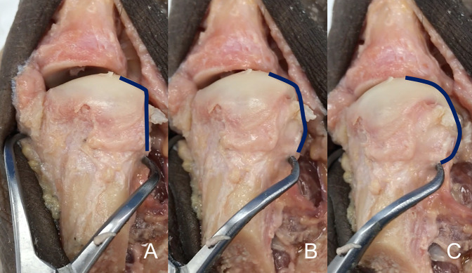
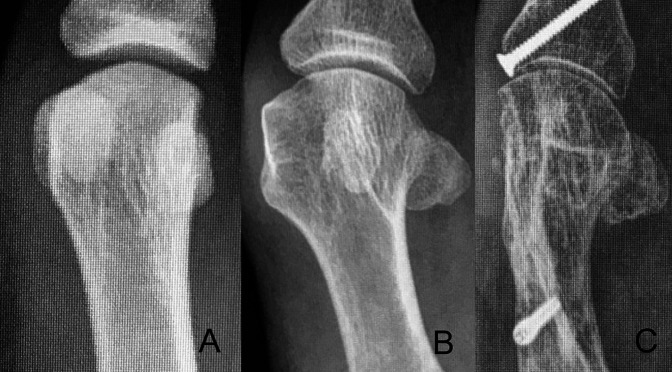
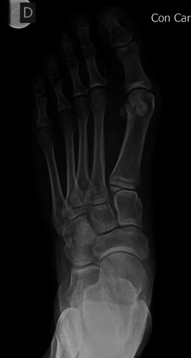
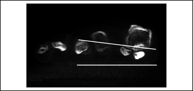
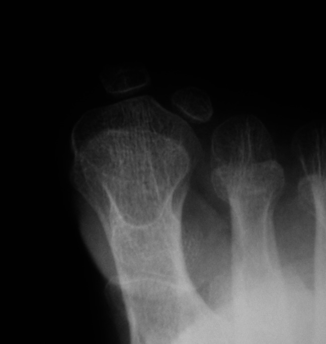
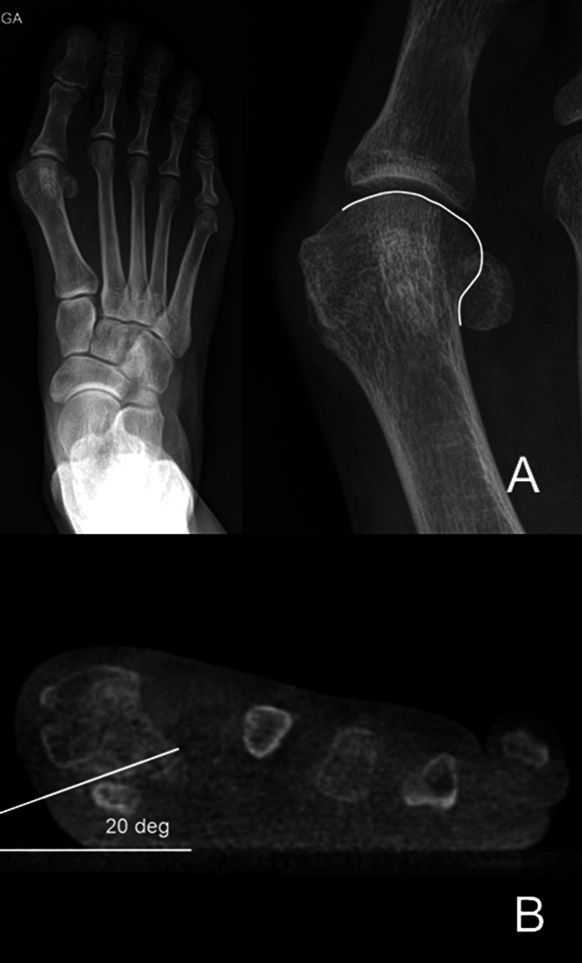
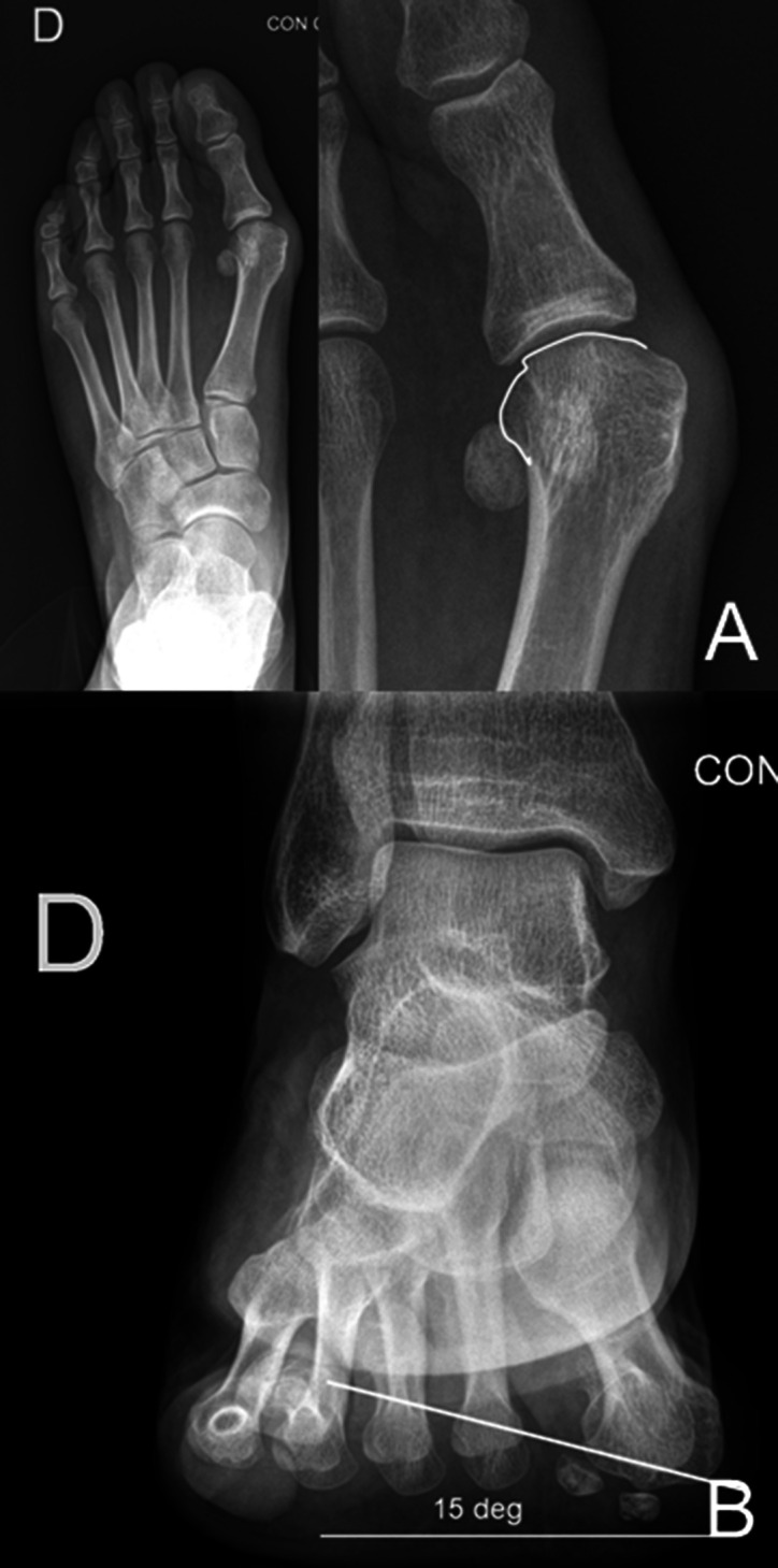
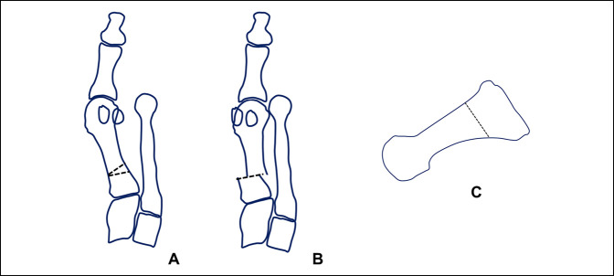
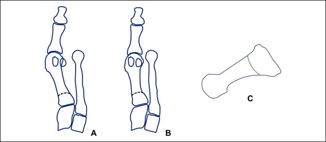
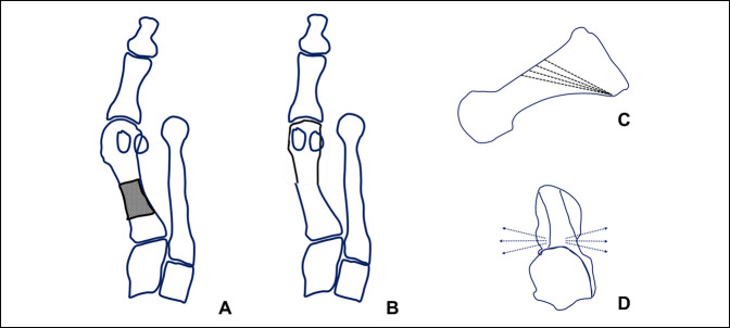
Hallux valgus deformity is a multiplanar deformity, where the rotational component has been recognized over the past 5 to 10 years and given considerable importance. Years ago, a rounded shape of the lateral edge of the first metatarsal head was identified as an important factor to detect after surgery because a less rounded metatarsal head was associated to less recurrence. More recently, pronation of the metatarsal bone was identified as the cause for the rounded appearance of the metatarsal head, and therefore, supination stress was found to be useful to achieve a better correction of the deformity. Using CT scans, up to 87% of hallux valgus cases have been shown to present with a pronated metatarsal bone, which highlights the multiplanar nature of the deformity. This pronation explained the perceived shape of the metatarsal bone and the malposition of the medial sesamoid bone in radiological studies, which has been associated as one of the most important factors for recurrence after treatment. Treatment options are discussed briefly, including metatarsal osteotomies and tarsometatarsal arthrodesis.

摘要

拇外翻畸形是一种多平面畸形,其中旋转成分在过去 5 到 10 年中得到了认可,并受到了相当大的重视。多年前,人们发现第一跖骨头外侧缘的圆形形状是术后检测的一个重要因素,因为较不圆的跖骨头与较低的复发率相关。最近,跖骨的内旋被认为是跖骨头呈圆形的原因,因此,旋前应力被发现对实现畸形的更好矫正很有用。使用 CT 扫描,高达 87%的拇外翻病例显示出跖骨的内旋,这突出了畸形的多平面性质。这种内旋解释了在影像学研究中所感知的跖骨形状和内侧籽骨的错位,这被认为是治疗后复发的最重要因素之一。简要讨论了治疗选择,包括跖骨截骨术和跗跖关节融合术。

https://cdn.ncbi.nlm.nih.gov/pmc/blobs/7abd/7322783/2b9945afb21d/jagrr-4-e20.00091-g012.jpg
https://cdn.ncbi.nlm.nih.gov/pmc/blobs/7abd/7322783/aab394e55adb/jagrr-4-e20.00091-g001.jpg
https://cdn.ncbi.nlm.nih.gov/pmc/blobs/7abd/7322783/d5a34dd29677/jagrr-4-e20.00091-g002.jpg
https://cdn.ncbi.nlm.nih.gov/pmc/blobs/7abd/7322783/a7bd616a98be/jagrr-4-e20.00091-g003.jpg
https://cdn.ncbi.nlm.nih.gov/pmc/blobs/7abd/7322783/d3817e2a5027/jagrr-4-e20.00091-g004.jpg
https://cdn.ncbi.nlm.nih.gov/pmc/blobs/7abd/7322783/5e8c694fdf43/jagrr-4-e20.00091-g005.jpg
https://cdn.ncbi.nlm.nih.gov/pmc/blobs/7abd/7322783/ecf591fdecc2/jagrr-4-e20.00091-g006.jpg
https://cdn.ncbi.nlm.nih.gov/pmc/blobs/7abd/7322783/aa679be69396/jagrr-4-e20.00091-g007.jpg
https://cdn.ncbi.nlm.nih.gov/pmc/blobs/7abd/7322783/c22c43ba5307/jagrr-4-e20.00091-g008.jpg
https://cdn.ncbi.nlm.nih.gov/pmc/blobs/7abd/7322783/b5d8e6982655/jagrr-4-e20.00091-g009.jpg
https://cdn.ncbi.nlm.nih.gov/pmc/blobs/7abd/7322783/0bd922db7899/jagrr-4-e20.00091-g010.jpg
https://cdn.ncbi.nlm.nih.gov/pmc/blobs/7abd/7322783/e1a6832550ac/jagrr-4-e20.00091-g011.jpg
https://cdn.ncbi.nlm.nih.gov/pmc/blobs/7abd/7322783/2b9945afb21d/jagrr-4-e20.00091-g012.jpg

相似文献

1
Metatarsal Pronation in Hallux Valgus Deformity: A Review.拇外翻畸形中的跖骨内翻:综述。
J Am Acad Orthop Surg Glob Res Rev. 2020 Jun 15;4(6). doi: 10.5435/JAAOSGlobal-D-20-00091. eCollection 2020 Jun.
2
Radiological and Anatomical Evaluation of First Metatarsal Pronation in Hallux Valgus Deformity: A Comparison Between Three Different Methods.拇外翻畸形中第一跖骨内旋的影像学和解剖学评估:三种不同方法的比较
J Foot Ankle Surg. 2023 May-Jun;62(3):448-454. doi: 10.1053/j.jfas.2022.11.002. Epub 2022 Nov 11.
3
Multiple factors contributing to the metatarsal head eversion in hallux valgus deformity. A prospective study using weight-bearing CT.导致拇外翻畸形中跖骨头外展的多种因素。一项使用负重 CT 的前瞻性研究。
Foot (Edinb). 2023 Dec;57:101965. doi: 10.1016/j.foot.2023.101965. Epub 2023 Feb 2.
4
Management of the Tarsometatarsal Joint in the Rotational Correction of Hallux Valgus by the Modified Lapidus Procedure: Intraoperative Technical Tips to Prevent Complications.改良Lapidus 手术矫正拇外翻时跗跖关节的处理:预防并发症的术中技术要点。
Foot Ankle Spec. 2021 Dec;14(6):528-533. doi: 10.1177/1938640020986686. Epub 2021 Jan 19.
5
Impact of First Metatarsal Hyperpronation on First Ray Alignment: A Study in Cadavers.第一跖骨过度旋前对第一跖骨排列的影响:尸体研究。
Clin Orthop Relat Res. 2022 Oct 1;480(10):2029-2040. doi: 10.1097/CORR.0000000000002265. Epub 2022 Jun 7.
6
Frontal Plane Rotation of the First Ray in Hallux Valgus using Standing Computerized Tomography (CT).第一跖骨在拇趾外翻中矢状面的旋转:使用站立式计算机断层扫描(CT)。
J Foot Ankle Surg. 2021 May-Jun;60(3):489-493. doi: 10.1053/j.jfas.2020.05.022. Epub 2020 Sep 21.
7
Axial rotation of the first metatarsal head in a normal population and hallux valgus patients.正常人群与足拇外翻患者第一跖骨头的轴向旋转。
Orthop Traumatol Surg Res. 2012 Oct;98(6):677-83. doi: 10.1016/j.otsr.2012.05.005. Epub 2012 Aug 28.
8
Intra‑ and Interobserver Reliability of Yamaguchi's Method for the Assessment of First Metatarsal Pronation in Hallux Valgus Deformity.山口法评估拇外翻畸形中第一跖骨内旋的观察者内及观察者间可靠性
J Foot Ankle Surg. 2023 May-Jun;62(3):432-436. doi: 10.1053/j.jfas.2022.10.008. Epub 2022 Oct 28.
9
A New Measure of Tibial Sesamoid Position in Hallux Valgus in Relation to the Coronal Rotation of the First Metatarsal in CT Scans.CT扫描中拇外翻时胫骨籽骨位置相对于第一跖骨冠状面旋转的一种新测量方法。
Foot Ankle Int. 2015 Aug;36(8):944-52. doi: 10.1177/1071100715576994. Epub 2015 Mar 26.
10
Coronal Plane Rotation of the Medial Column in Hallux Valgus: A Retrospective Case-Control Study.拇外翻中内侧柱冠状面旋转:一项回顾性病例对照研究。
Foot Ankle Int. 2022 Aug;43(8):1041-1048. doi: 10.1177/10711007221091810. Epub 2022 May 2.

引用本文的文献

1
Reliability of categorization of the shape of the lateral edge of the first metatarsal head in hallux valgus.拇外翻中第一跖骨头外侧缘形状分类的可靠性
Medicine (Baltimore). 2025 Jul 18;104(29):e43203. doi: 10.1097/MD.0000000000043203.
2
Artificial intelligence-based diagnosis of hallux valgus interphalangeus using anteroposterior foot radiographs.基于人工智能利用足部正位X线片诊断踇趾中间关节外翻
World J Orthop. 2025 Jun 18;16(6):103832. doi: 10.5312/wjo.v16.i6.103832.
3
Comparative Analysis of Minimally Invasive and Open Proximal Chevron-Akin Osteotomies in Moderate-to-Severe Hallux Valgus Deformity.

本文引用的文献

1
Republication of "Proximal Rotational Metatarsal Osteotomy for Hallux Valgus (PROMO): Short-term Prospective Case Series With a Novel Technique and Topic Review".《拇外翻近端旋转跖骨截骨术(PROMO):采用新技术的短期前瞻性病例系列及专题综述》再版
Foot Ankle Orthop. 2023 Aug 14;8(3):24730114231195049. doi: 10.1177/24730114231195049. eCollection 2023 Jul.
2
The shape of the first metatarsal head and its association with the presence of sesamoid-metatarsal joint osteoarthritis and the pronation angle.第一跖骨头的形状及其与籽骨-跖骨关节骨关节炎的存在和内旋角度的关联。
J Orthop Sci. 2020 Jul;25(4):658-663. doi: 10.1016/j.jos.2019.06.013. Epub 2019 Jul 17.
3
微创与开放式近端 Chevron-Akin 截骨术治疗中重度拇外翻畸形的对比分析
Clin Orthop Surg. 2025 Jun;17(3):514-522. doi: 10.4055/cios24450. Epub 2025 Apr 18.
4
Sesamoid correction achieved during the learning curve for Scarf-Akin osteotomy without lateral soft-tissue release: a single-centre prospective observational study.在不进行外侧软组织松解的Scarf-Akin截骨术学习曲线期间实现籽骨矫正:一项单中心前瞻性观察研究。
Arch Orthop Trauma Surg. 2025 May 9;145(1):284. doi: 10.1007/s00402-025-05883-z.
5
Double osteotomy in recurrence cases with distal metatarsal articular angle increase after hallux valgus distal surgery.拇外翻远端手术后远端跖骨关节角增大的复发病例中的双截骨术
Jt Dis Relat Surg. 2025 Apr 10;36(2):420-427. doi: 10.52312/jdrs.2025.2161.
6
Weight-Bearing CT: Advancing the Diagnosis and Treatment of Hallux Valgus, Midfoot Pathology, and Progressive Collapsing Foot Deformity.负重CT:推进拇外翻、中足病变及进行性足部塌陷畸形的诊断与治疗
Diagnostics (Basel). 2025 Jan 31;15(3):343. doi: 10.3390/diagnostics15030343.
7
Assessing the AIR classification reliability for estimating pronation of the first metatarsal.评估用于估计第一跖骨旋前的AIR分类可靠性。
BMC Musculoskelet Disord. 2024 Dec 19;25(1):1034. doi: 10.1186/s12891-024-08158-7.
8
Revisiting the radiological signs for the first metatarsal pronation assessment.重新审视用于第一跖骨内旋评估的影像学征象。
Bone Jt Open. 2024 Nov 15;5(11):1037-1040. doi: 10.1302/2633-1462.511.BJO-2024-0116.R1.
9
Effects of Pathological Characteristics on Functional Scores and Surgical Satisfaction in Patients after Hallux Valgus Surgery.病理特征对拇外翻手术后患者功能评分和手术满意度的影响。
Orthop Surg. 2024 Dec;16(12):2984-2994. doi: 10.1111/os.14237. Epub 2024 Sep 18.
10
Preoperative Degenerative Changes at the Tibial Sesamoid-Metatarsal Joint in Hallux Valgus: Association With Postoperative Patient-Reported Outcomes After Modified Lapidus Procedure.拇外翻患者胫侧籽骨-跖骨关节术前的退变改变:与改良Lapidus手术后患者报告的术后结果的关联
Foot Ankle Orthop. 2024 Jun 3;9(2):24730114241256370. doi: 10.1177/24730114241256370. eCollection 2024 Apr.
Effect of natural full weight-bearing during standing on the rotation of the first metatarsal bone.
自然全负重站立对第一跖骨旋转的影响。
Clin Anat. 2019 Jul;32(5):715-721. doi: 10.1002/ca.23385. Epub 2019 Apr 29.
4
Importance of postoperative sesamoid reduction on the outcomes of proximal chevron osteotomy for moderate to severe hallux valgus deformity.重视术后籽骨复位在中重度拇外翻畸形行近端Chevron 截骨术后的疗效。
Foot Ankle Surg. 2019 Aug;25(4):434-440. doi: 10.1016/j.fas.2018.02.006. Epub 2018 Feb 16.
5
Pilot Study of a 3-Dimensional Method for Analysis of Pronation of the First Metatarsal of Hallux Valgus Patients.第一跖骨内翻的拇外翻患者旋前三维分析的初步研究。
Foot Ankle Int. 2018 Dec;39(12):1449-1456. doi: 10.1177/1071100718793391. Epub 2018 Sep 11.
6
Sesamoid Position in Hallux Valgus in Relation to the Coronal Rotation of the First Metatarsal.拇外翻中籽骨位置与第一跖骨冠状面旋转的关系
Foot Ankle Clin. 2018 Jun;23(2):219-230. doi: 10.1016/j.fcl.2018.01.003.
7
Is the Rotational Deformity Important in Our Decision-Making Process for Correction of Hallux Valgus Deformity?在我们矫正拇外翻畸形的决策过程中,旋转畸形重要吗?
Foot Ankle Clin. 2018 Jun;23(2):205-217. doi: 10.1016/j.fcl.2018.01.009. Epub 2018 Mar 9.
8
Factors Associated With Early Loss of Hallux Valgus Correction.与拇外翻矫正早期失败相关的因素
J Foot Ankle Surg. 2018 Mar-Apr;57(2):236-240. doi: 10.1053/j.jfas.2017.08.018. Epub 2017 Dec 15.
9
Recurrence of Hallux Valgus Can Be Predicted from Immediate Postoperative Non-Weight-Bearing Radiographs.拇外翻复发可通过术后即刻非负重位X线片预测。
J Bone Joint Surg Am. 2017 Jul 19;99(14):1190-1197. doi: 10.2106/JBJS.16.00980.
10
Rotational Osteotomy for Hallux Valgus. A New Technique for Primary and Revision Cases.拇外翻旋转截骨术。一种用于初次手术及翻修病例的新技术。
Tech Foot Ankle Surg. 2017 Mar;16(1):3-10. doi: 10.1097/BTF.0000000000000142. Epub 2016 Nov 17.