Department of Orthopaedics and Trauma Surgery, University Hospital Bonn, Germany.
Department of Anesthesiology and Intensive Care Medicine, University Hospital Bonn, Germany.
Oxid Med Cell Longev. 2020 May 14;2020:7606938. doi: 10.1155/2020/7606938. eCollection 2020.
Myocardial ischemia and reperfusion (I/R) injury is associated with oxidative stress and inflammation, leading to scar development and malfunction. The marine omega-3 fatty acids (-3 FA), eicosapentaenoic acid (EPA), and docosahexaenoic acid (DHA) are mediating cardioprotection and improving clinical outcomes in patients with heart disease. Therefore, we tested the hypothesis that docosahexaenoic acid (DHA) supplementation prior to LAD occlusion-induced myocardial injury (MI) confers cardioprotection in mice.
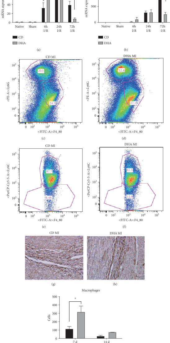
C57BL/6N mice were placed on DHA or control diets (CD) beginning 7 d prior to 60 min LAD occlusion-induced MI or sham surgery. The expression of inflammatory mediators was measured via RT-qPCR. Besides FACS analysis for macrophage quantification and subtype evaluation, macrophage accumulation as well as collagen deposition was quantified in histological sections. Cardiac function was assessed using a pressure-volume catheter for up to 14 d.
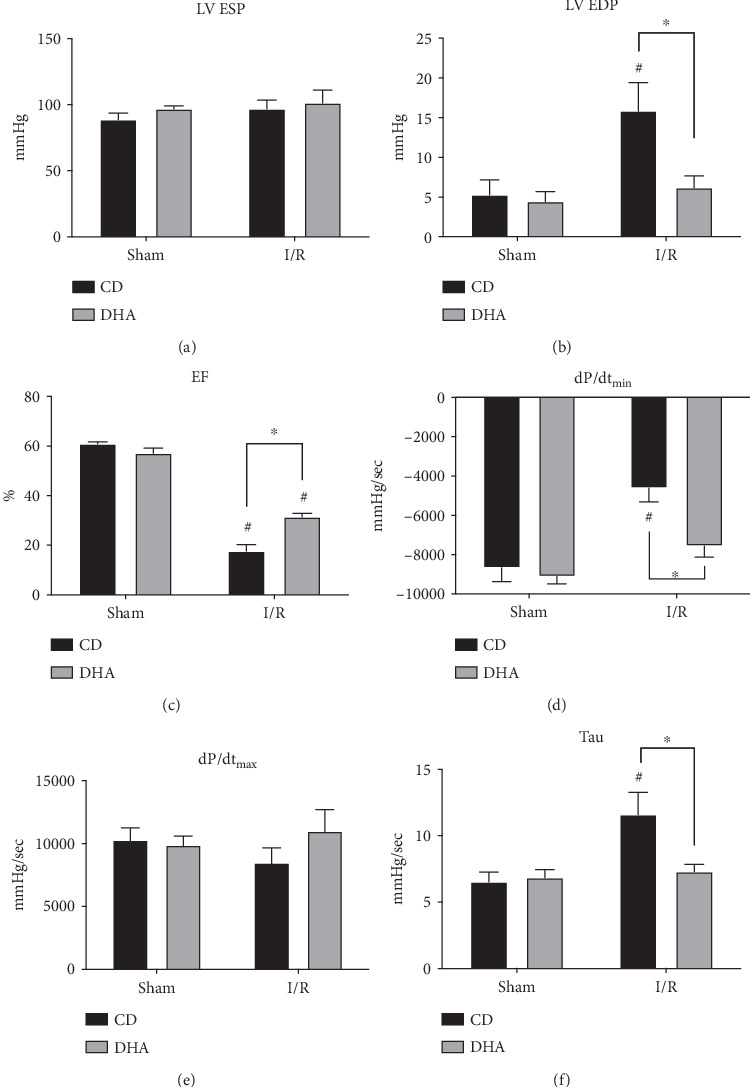
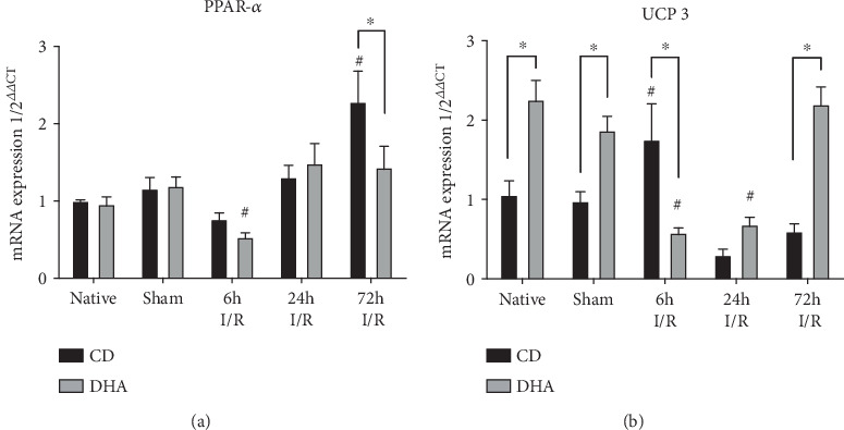
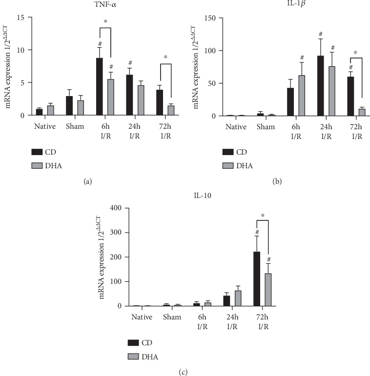
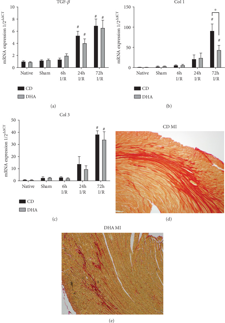
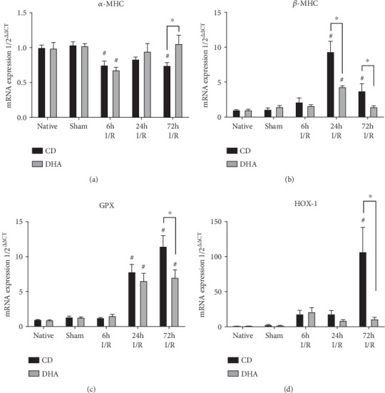
DHA supplementation significantly attenuated the induction of peroxisome proliferator-activated receptor- (PPAR-) (2.3 ± 0.4 CD vs. 1.4 ± 0.3 DHA) after LAD occlusion. Furthermore, TNF- (4.0 ± 0.6 CD vs. 1.5 ± 0.2 DHA), IL-1 (60.7 ± 7.0 CD vs. 11.6 ± 1.9 DHA), and IL-10 (223.8 ± 62.1 CD vs. 135.5 ± 38.5 DHA) mRNA expression increase was diminished in DHA-supplemented mice after 72 h reperfusion. These changes were accompanied by a less prominent switch in / myosin heavy chain isoforms. Chemokine mRNA expression was stronger initiated (CCL2 6 h: 32.8 ± 11.5 CD vs. 78.8 ± 13.6 DHA) but terminated earlier (CCL2 72 h: 39.5 ± 7.8 CD vs. 8.2 ± 1.9 DHA; CCL3 72 h: 794.3 ± 270.9 CD vs. 258.2 ± 57.8 DHA) in DHA supplementation compared to CD mice after LAD occlusion. Correspondingly, DHA supplementation was associated with a stronger increase of predominantly alternatively activated Ly6C-positive macrophage phenotype, being associated with less collagen deposition and better LV function (EF 14 d: 17.6 ± 2.6 CD vs. 31.4 ± 1.5 DHA).
Our data indicate that DHA supplementation mediates cardioprotection from MI via modulation of the inflammatory response with timely and attenuated remodeling. DHA seems to attenuate MI-induced cardiomyocyte injury partly by transient PPAR- downregulation, diminishing the need for antioxidant mechanisms including mitochondrial function, or - to -MHC isoform switch.
心肌缺血再灌注(I/R)损伤与氧化应激和炎症有关,导致瘢痕形成和功能障碍。海洋 ω-3 脂肪酸(-3 FA),二十碳五烯酸(EPA)和二十二碳六烯酸(DHA)可介导心脏保护作用,并改善心脏病患者的临床结局。因此,我们检验了这样一个假设,即在左前降支闭塞引起心肌损伤(MI)之前补充二十二碳六烯酸(DHA)可赋予小鼠心脏保护作用。
C57BL/6N 小鼠在左前降支闭塞诱导的 MI 或假手术后 7 天开始接受 DHA 或对照饮食(CD)。通过 RT-qPCR 测量炎症介质的表达。除了用于巨噬细胞定量和亚型评估的 FACS 分析外,还在组织学切片中定量了巨噬细胞积聚和胶原沉积。使用压力-容积导管评估心功能长达 14 天。
DHA 补充可显著抑制左前降支闭塞后过氧化物酶体增殖物激活受体-(PPAR-)(2.3 ± 0.4 CD 与 1.4 ± 0.3 DHA)的诱导。此外,TNF-(4.0 ± 0.6 CD 与 1.5 ± 0.2 DHA),IL-1(60.7 ± 7.0 CD 与 11.6 ± 1.9 DHA)和 IL-10(223.8 ± 62.1 CD 与 135.5 ± 38.5 DHA)mRNA 表达在再灌注 72 小时后在 DHA 补充的小鼠中减少。这些变化伴随着 / 肌球蛋白重链同工型的变化不明显。趋化因子 mRNA 表达更早启动(CCL2 6 h:32.8 ± 11.5 CD 与 78.8 ± 13.6 DHA)但更早终止(CCL2 72 h:39.5 ± 7.8 CD 与 8.2 ± 1.9 DHA; CCL3 72 h:794.3 ± 270.9 CD 与 258.2 ± 57.8 DHA)在左前降支闭塞后 DHA 补充的小鼠中。相应地,DHA 补充与主要以替代激活 Ly6C 阳性巨噬细胞表型为主的更强增加有关,与胶原沉积较少和更好的 LV 功能(EF 14 d:17.6 ± 2.6 CD 与 31.4 ± 1.5 DHA)有关。
我们的数据表明,DHA 补充通过调节炎症反应来介导 MI 的心脏保护作用,从而实现及时和减弱的重塑。DHA 似乎通过瞬时 PPAR-下调来减轻 MI 诱导的心肌细胞损伤,减少对包括线粒体功能在内的抗氧化机制的需求,或者向-MHC 同工型转换。