Wang Tao, Zhong Lei, Yin Shiyi, Bao Tiancheng, Yang Jiezheng, Wang Ting, Ling Shiqi
Department of Ophthalmology, Third Affiliated Hospital of Sun Yat-Sen University, Guangzhou, Guangdong, China.
State Key Laboratory of Ophthalmology, Zhongshan Ophthalmic Center, Sun Yat-sen University, Guangzhou, Guangdong, China.
J Ophthalmol. 2020 Aug 11;2020:9108317. doi: 10.1155/2020/9108317. eCollection 2020.
The present study highlighted the value of anterior segment optical coherence tomography (AS-OCT) for different types of corneal foreign bodies in humans.
This study was a prospective observational study. The patients included were divided into two groups. If the patients were directly diagnosed based on eye injury history and slit-lamp examination, then they were assigned to Group A. Otherwise, the patients were assigned to Group B. We compared and described the characteristics of the corneal foreign body in both groups using AS-OCT.
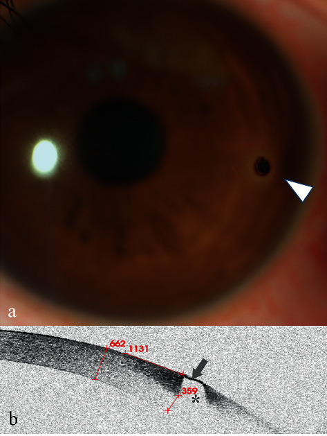
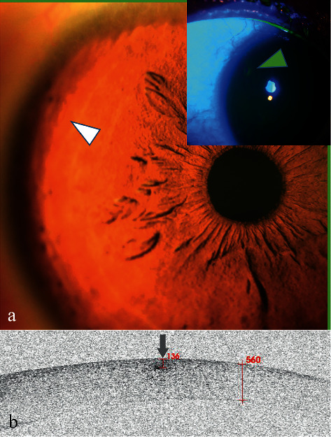
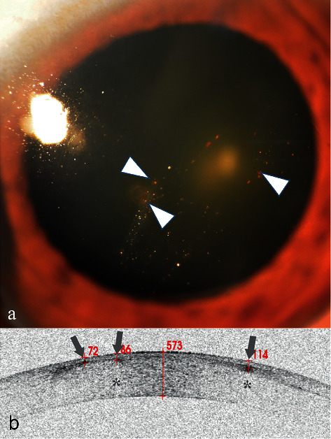
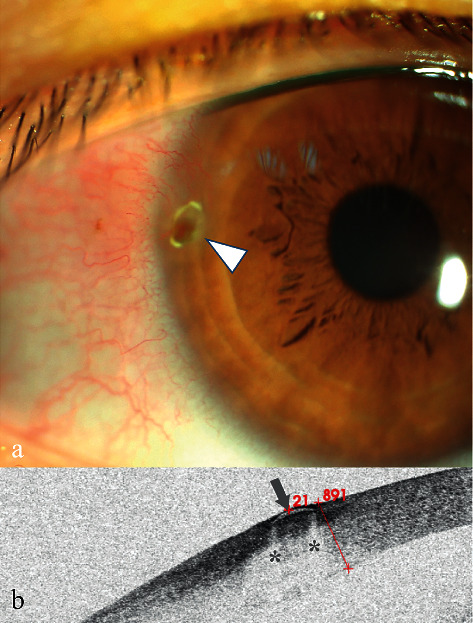
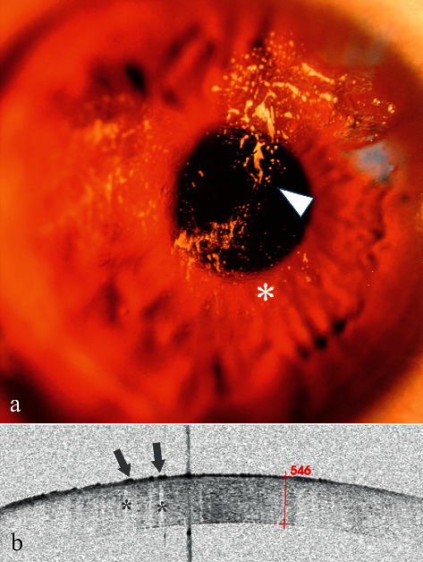
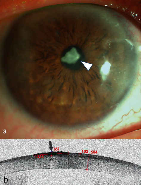
From October 2017 to January 2020, 36 eyes of 36 patients (9 females and 27 males) with a mean age of 37.8 ± 11.7 years were included in the study. Patients in Group A were the majority and accounted for 72.2% (26/36). High signals on AS-OCT images were the main constituent and accounted for 92.3% (24/26) in Group A and 70.0% (7/10) in Group B. Most of the patients in Group A, 96.2% (25/26), had clear boundaries. A blurred boundary was observed in 70.0% (7/10) of the patients in Group B. The foreign bodies on AS-OCT images had key characteristics of a high signal followed by a central zone shadowing effect and a low signal followed by a marginal zone shadowing effect. Further, all of the lesions could be directly located in Group B, and 92.3% (24/26) of the patients in Group A did not have directly located lesions. Six representative cases are described in detail.
AS-OCT is a valuable tool in the diagnosis and management of corneal foreign bodies, especially for unusual corneal foreign body.
本研究强调了眼前节光学相干断层扫描(AS-OCT)在诊断人类不同类型角膜异物中的价值。
本研究为前瞻性观察性研究。纳入的患者分为两组。若患者根据眼外伤史和裂隙灯检查直接确诊,则归入A组。否则,患者归入B组。我们使用AS-OCT比较并描述了两组角膜异物的特征。
2017年10月至2020年1月,本研究纳入了36例患者的36只眼(9例女性,27例男性),平均年龄为37.8±11.7岁。A组患者占大多数,占72.2%(26/36)。AS-OCT图像上的高信号是主要成分,在A组中占92.3%(24/26),在B组中占70.0%(7/10)。A组大多数患者,96.2%(25/26),边界清晰。B组70.0%(7/10)的患者观察到边界模糊。AS-OCT图像上的异物具有高信号后伴中央区阴影效应以及低信号后伴边缘区阴影效应的关键特征。此外,B组所有病变均可直接定位,A组92.3%(24/26)的患者病变无法直接定位。详细描述了6个代表性病例。
AS-OCT是诊断和处理角膜异物的一种有价值的工具,尤其是对于不常见的角膜异物。