Department of Orthopaedics, Chinese People's Liberation Army General Hospital, 28 Fuxing Road, Haidian District, Beijing, China 100853.
Biomed Res Int. 2020 Aug 25;2020:4809013. doi: 10.1155/2020/4809013. eCollection 2020.
The purpose of this study was to establish the finite element analysis (FEA) model of acetabular bone defect in Crowe type II or III developmental dysplasia of the hip (DDH), which could evaluate the stability of the acetabular cup with different types of bone defects, different diameters of femoral ceramic heads, and the use of screws and analyze the stress distribution of screws.
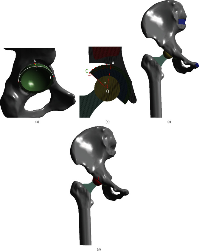
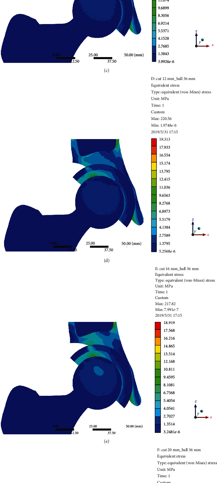
The FEA model was based on the CT scan of a female patient without any acetabular bone defect. The model of acetabular bone defect in total hip arthroplasty for Crowe II or III DDH was made by the increasing superolateral bone defect area of the acetabular cup. Point A was located in the most medial part of the acetabular bone defect. A 52 mm PINNACLE cup with POROCOAT Porous coating was implanted, and two screws (the lengths were 25 mm and 40 mm) were implanted to fix the acetabular cup. The stability of the acetabular cup and the von Mises stress of point A and screws were analyzed by a single-legged stance loading applied in 1948 N (normal working). The different diameters of the femoral ceramic head (28 mm, 32 mm, and 36 mm) were also analyzed.
The von Mises stress of point A was gradually increased with the increasing uncoverage values. When the uncoverage values exceeded 24.5%, the von Mises stress of point A without screws increased significantly, leading to instability of the cup. Screws could effectively reduce the von Mises stress of point A with uncoverage values of more than 24.5%. However, the peak von Mises stress in the screws with the uncoverage values that exceeded 24.5% was considerably increased. The diameter of the femoral ceramic head had no significant effect on the von Mises stress and the stability of the acetabular cup.
We recommend that uncoverage values of less than 24.5% with or without screw is safe for patients with Crowe II or III DDH.
本研究旨在建立 Crowe Ⅱ或Ⅲ型发育性髋关节发育不良(DDH)髋臼骨缺损的有限元分析(FEA)模型,以评估不同类型髋臼骨缺损、不同直径股骨陶瓷头、使用螺钉的髋臼杯稳定性,并分析螺钉的应力分布。
FEA 模型基于一位无髋臼骨缺损的女性患者的 CT 扫描。通过增加髋臼杯的超外侧骨缺损面积来制作 Crowe II 或 III DDH 全髋关节置换术的髋臼骨缺损模型。点 A 位于髋臼骨缺损最内侧部分。植入 52mm PINNACLE 杯,带有 POROCOAT 多孔涂层,并植入两根螺钉(长度分别为 25mm 和 40mm)以固定髋臼杯。通过单腿站立施加 1948N(正常工作)的负荷来分析髋臼杯的稳定性和点 A 及螺钉的 von Mises 应力。还分析了不同直径的股骨陶瓷头(28mm、32mm 和 36mm)。
点 A 的 von Mises 应力随覆盖值的增加而逐渐增加。当覆盖值超过 24.5%时,无螺钉的点 A 的 von Mises 应力显著增加,导致杯不稳定。螺钉可有效降低覆盖值超过 24.5%的点 A 的 von Mises 应力。然而,覆盖值超过 24.5%时,螺钉的 von Mises 峰值应力显著增加。股骨陶瓷头的直径对髋臼杯的 von Mises 应力和稳定性没有显著影响。
我们建议 Crowe Ⅱ或Ⅲ型 DDH 患者的覆盖值小于 24.5%(有或无螺钉)是安全的。