The Thoracic Surgery Oncology Laboratory and the International Mesothelioma Program (www.impmeso.org), Division of Thoracic Surgery and the Lung Center, Brigham and Women's Hospital, and Harvard Medical School, Boston, MA, USA.
Department of Pathology, Brigham and Women's Hospital, and Harvard Medical School, Boston, MA, USA.
J Pathol. 2021 Jan;253(1):68-79. doi: 10.1002/path.5551. Epub 2020 Oct 15.
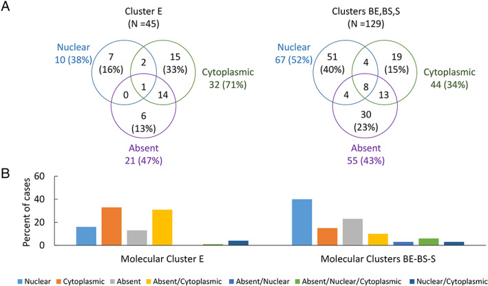
BRCA1-associated protein-1 (BAP1) expression is commonly lost in several tumors including malignant pleural mesothelioma (MPM). Presence or absence of immunohistochemical BAP1 nuclear staining in tumor cells is currently used for differential diagnosis of MPM. In this study, a large cohort of 596 MPM tumors with available clinical data was analyzed to examine associations of BAP1 staining pattern with clinical and molecular features that may reflect the impact of BAP1 mutation on MPM biology. Cases were classified according to the BAP1 staining pattern of tumor cells. Exome and RNA-sequencing data were available for subsets of cases. Levels of mRNA encoding claudin 15 (CLDN15) and vimentin (VIM) were determined using RT-qPCR on 483 cases to estimate the relative proportions of epithelial-like and mesenchymal-like components in each tumor. Four BAP1 staining patterns were observed: single-pattern nuclear staining (36%), single-pattern cytoplasmic staining (25%), single-pattern absent staining (12%), and combinations of these staining patterns (27%). This study confirmed prior reports that nuclear BAP1 is more frequently associated with wild-type BAP1 and sarcomatoid histology. However, no associations between BAP1 staining pattern(s) and mutations in specific protein domains and/or mutation type were observed. BAP1 staining patterns were significantly associated (p < 0.001) with BAP1 gene expression, MPM histologic subtypes, molecular clusters, and markers of epithelial-to-mesenchymal transition. Frequent observation of combinations of BAP1 staining patterns in MPM tumors indicated intra-tumoral heterogeneity of BAP1 status. Cytoplasmic BAP1 staining was identified as a putative indicator of favorable prognosis in non-epithelioid MPM. In conclusion, novel significant associations among different BAP1 staining patterns and subgroups of MPM tumors were observed, suggesting that the role of BAP1 in tumor progression may be more complex than its presumed tumor suppressor function. Cytoplasmic staining was identified as a putative indicator of favorable prognosis in non-epithelioid MPM, potentially addressing a critical need in clinical decision-making in this disease. © 2020 The Authors. The Journal of Pathology published by John Wiley & Sons, Ltd. on behalf of The Pathological Society of Great Britain and Ireland.
BRCA1 相关蛋白-1(BAP1)表达在包括恶性胸膜间皮瘤(MPM)在内的几种肿瘤中通常丢失。肿瘤细胞中免疫组化 BAP1 核染色的存在或缺失目前用于 MPM 的鉴别诊断。在这项研究中,对 596 例具有可用临床数据的 MPM 肿瘤进行了大样本分析,以研究 BAP1 染色模式与可能反映 BAP1 突变对 MPM 生物学影响的临床和分子特征之间的关联。根据肿瘤细胞的 BAP1 染色模式对病例进行分类。部分病例可提供外显子和 RNA 测序数据。在 483 例病例中使用 RT-qPCR 测定编码紧密连接蛋白 15(CLDN15)和波形蛋白(VIM)的 mRNA 水平,以估计每个肿瘤中上皮样和间充质样成分的相对比例。观察到四种 BAP1 染色模式:单一核染色模式(36%)、单一胞质染色模式(25%)、单一无染色模式(12%)和这些染色模式的组合(27%)。本研究证实了先前的报道,即核 BAP1 更常与野生型 BAP1 和肉瘤样组织学相关。然而,未观察到 BAP1 染色模式与特定蛋白结构域和/或突变类型的突变之间存在关联。BAP1 染色模式与 BAP1 基因表达、MPM 组织学亚型、分子簇和上皮-间充质转化标志物显著相关(p<0.001)。在 MPM 肿瘤中频繁观察到 BAP1 染色模式的组合表明 BAP1 状态存在肿瘤内异质性。细胞质 BAP1 染色被确定为非上皮样 MPM 中有利预后的潜在指标。总之,观察到不同的 BAP1 染色模式和 MPM 肿瘤亚组之间存在新的显著关联,表明 BAP1 在肿瘤进展中的作用可能比其假定的肿瘤抑制功能更为复杂。细胞质染色被确定为非上皮样 MPM 中有利预后的潜在指标,可能解决了该疾病临床决策中的关键需求。2020 年,作者。约翰威立父子公司代表英国和爱尔兰病理学学会出版的《病理学杂志》。