Institute of Physiology, Justus-Liebig-University Giessen, Aulweg 129, 35392, Gießen, Germany.
Basic Res Cardiol. 2020 Nov 10;115(6):65. doi: 10.1007/s00395-020-00824-w.
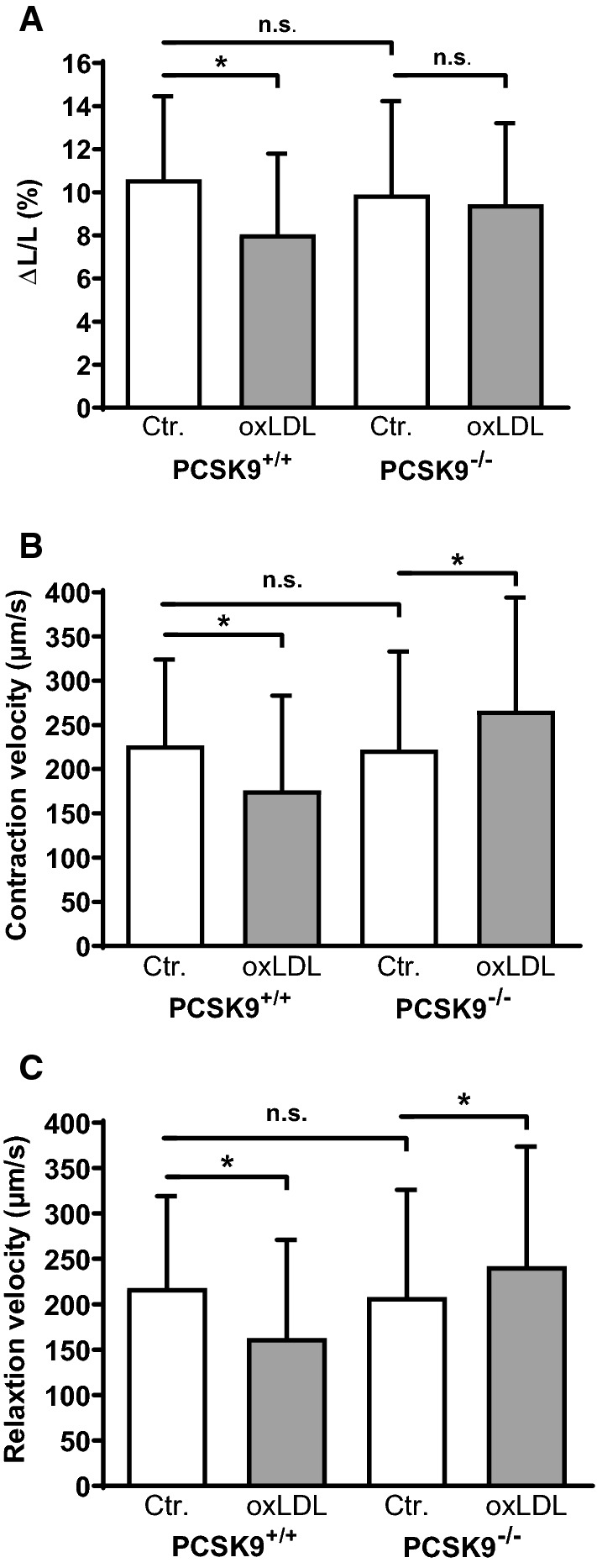
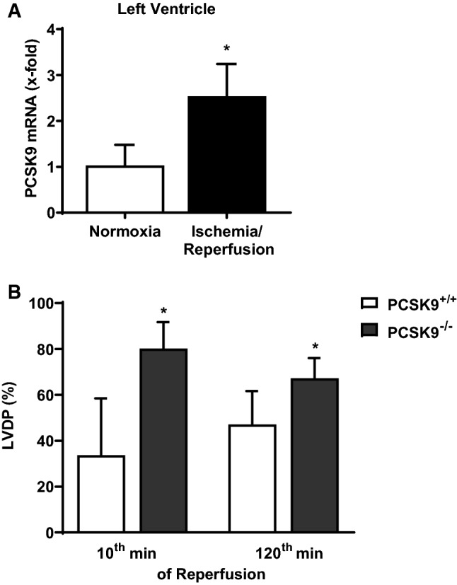
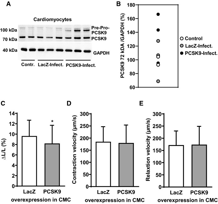
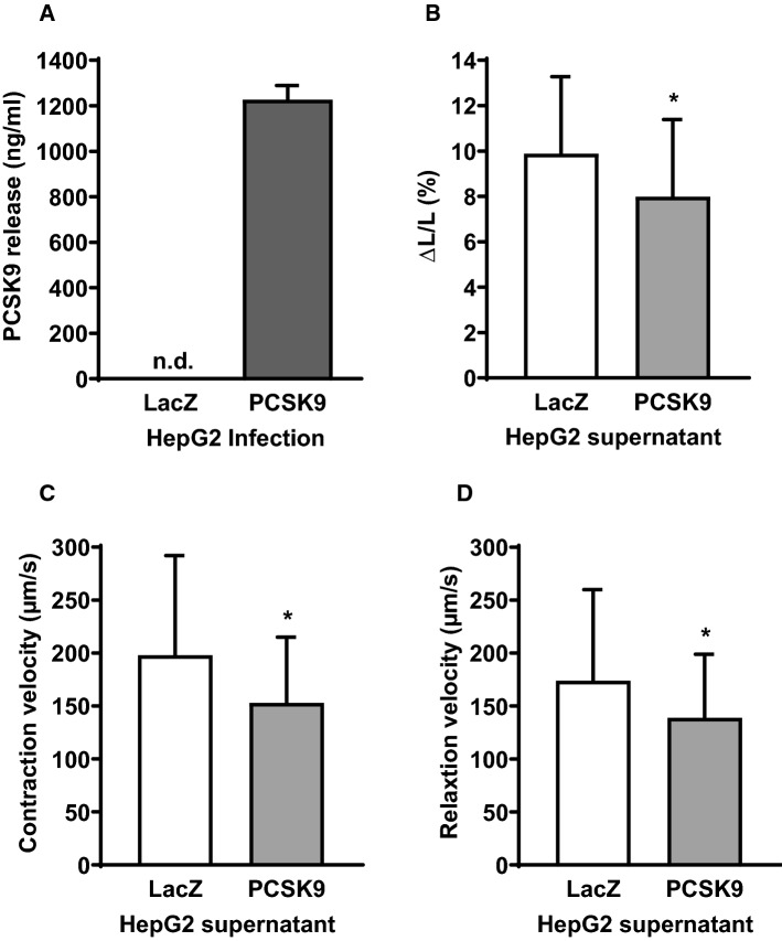
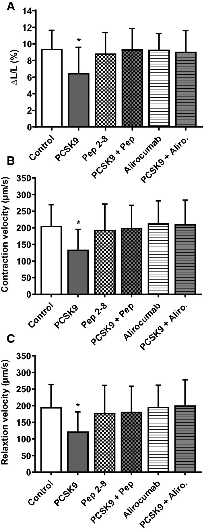
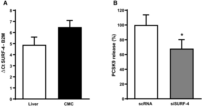
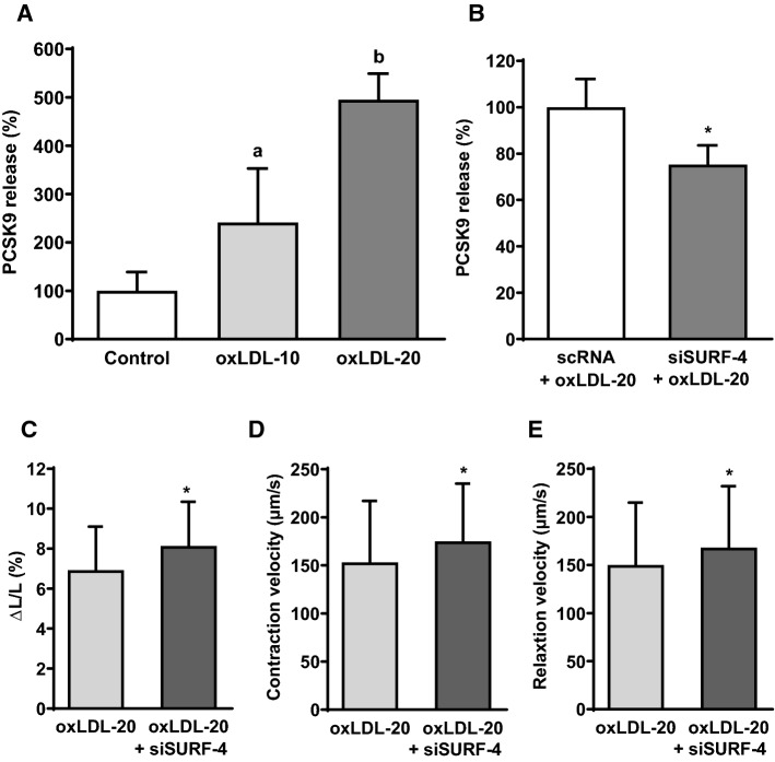
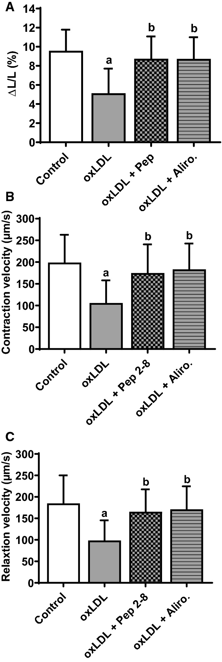
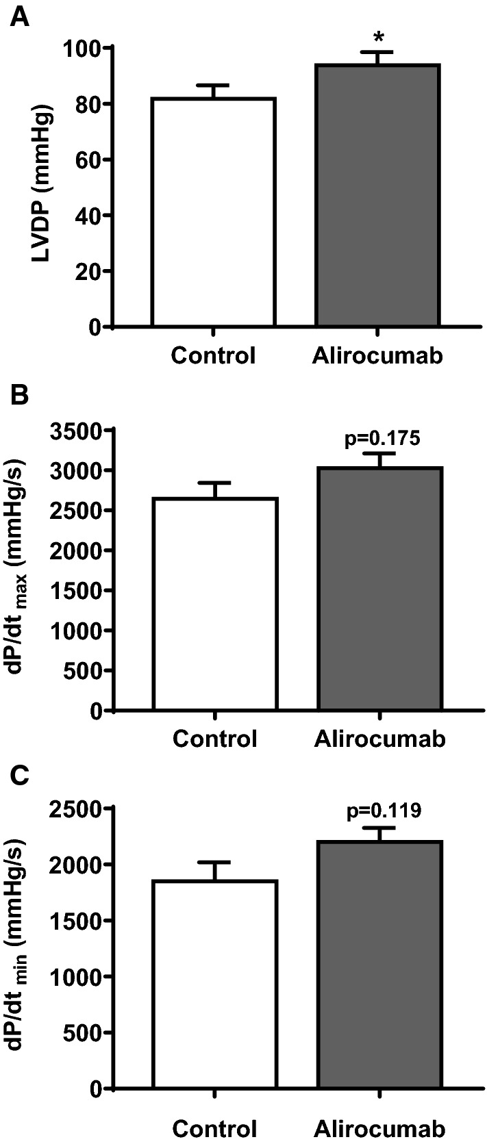
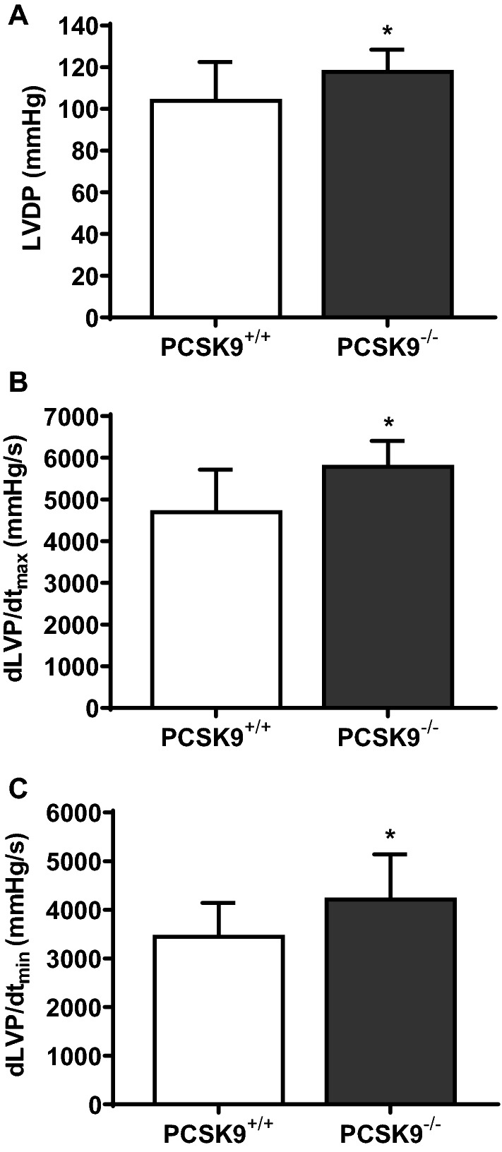
Proprotein convertase subtilisin kexin type 9 (PCSK9) is in the focus of cardiovascular research due to its role in hepatic low density lipoprotein (LDL) clearance. However, extrahepatic expression of PCSK9 such as in cardiomyocytes and its regulation by oxidized LDL (oxLDL) put notion on extrahepatic effects of PCSK9 as well. This study was aimed to reveal the role of PCSK9 in oxLDL-dependent regulation of cardiomyocyte function. Adult rat and mouse ventricular cardiomyocytes and isolated perfused hearts were used. OxLDL was applied to increase PCSK9 expression in cardiomyocytes. Cell function was analyzed by load-free cell shortening as well as left ventricular developed pressure of isolated hearts. OxLDL decreased shortening in wild-type-derived mouse cardiomyocytes but not in those isolated from PCSK9 knockout mice. Overexpression of human PCSK9 in rat cardiomyocytes reduced shortening in the absence of oxLDL. Addition of recombinant PCSK9 mimicked these effects. In cardiomyocytes, oxLDL induced PCSK9 release into the supernatant. Inhibition of PCSK9 by Pep 2-8 or alirocumab attenuated the oxLDL-induced loss of cardiomyocyte shortening. Cardiomyocytes express surfeit locus protein 4 (SURF-4), a protein required for PCSK9 secretion in human embryonic kidney cells (HEK 293 T), and silencing of SURF-4 reduced the oxLDL effects on cardiomyocytes. In isolated perfused rat hearts PCSK9 inhibition by alirocumab improved the function. In addition, left ventricular function of isolated hearts from PCSK9 knockout mice was increased under basal conditions as well as at 10 min and 120 min of reperfusion following 45 min of ischemia. Collectively, the data show that cardiomyocytes express and release PCSK9 that acts in an autocrine way on cardiomyocytes and impairs their function.
前蛋白转化酶枯草溶菌素 9(PCSK9)在肝脏低密度脂蛋白(LDL)清除中起作用,因此成为心血管研究的焦点。然而,PCSK9 在心肌细胞中的表达以及氧化型 LDL(oxLDL)对其的调节提示了 PCSK9 的非肝脏作用。本研究旨在揭示 PCSK9 在 oxLDL 依赖性调节心肌细胞功能中的作用。使用成年大鼠和小鼠心室心肌细胞以及分离的灌注心脏。应用 oxLDL 增加心肌细胞中的 PCSK9 表达。通过无负荷细胞缩短以及分离心脏的左心室发展压分析细胞功能。oxLDL 降低了野生型衍生的小鼠心肌细胞的缩短,但不降低来自 PCSK9 敲除小鼠的心肌细胞的缩短。在大鼠心肌细胞中过表达人 PCSK9 在没有 oxLDL 的情况下减少了缩短。添加重组 PCSK9 模拟了这些作用。在心肌细胞中,oxLDL 将 PCSK9 释放到上清液中。用 Pep 2-8 或 alirocumab 抑制 PCSK9 减弱了 oxLDL 诱导的心肌细胞缩短丧失。心肌细胞表达过剩座位蛋白 4(SURF-4),这是人类胚胎肾细胞(HEK 293T)中 PCSK9 分泌所必需的蛋白,沉默 SURF-4 减少了 oxLDL 对心肌细胞的作用。在分离的灌注大鼠心脏中,用 alirocumab 抑制 PCSK9 改善了功能。此外,在缺血 45 分钟后,10 分钟和 120 分钟再灌注期间,PCSK9 敲除小鼠的分离心脏的左心室功能增加。总之,这些数据表明心肌细胞表达和释放 PCSK9,该蛋白以自分泌方式作用于心肌细胞并损害其功能。