Chen Dayong, Qiao Hai, Wang Yiting, Yin Na, Fang Liaoqiong, Wang Zhibiao
State Key Laboratory of Ultrasound Engineering in Medicine Co-Founded by Chongqing and the Ministry of Science and Technology, College of Biomedical Engineering, Chongqing Key Laboratory of Biomedical Engineering; Chongqing Medical University, 40016, PR China.
Genes Dis. 2020 Jan 23;7(4):636-648. doi: 10.1016/j.gendis.2020.01.011. eCollection 2020 Dec.
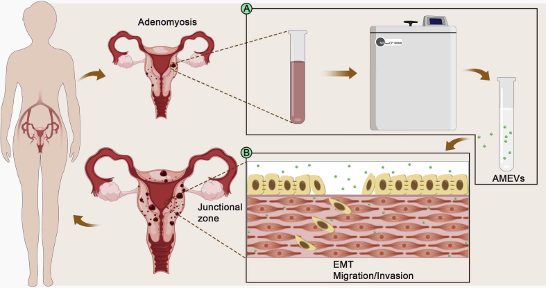
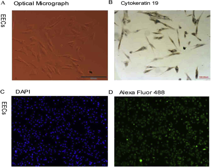
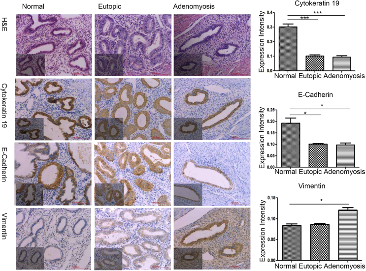
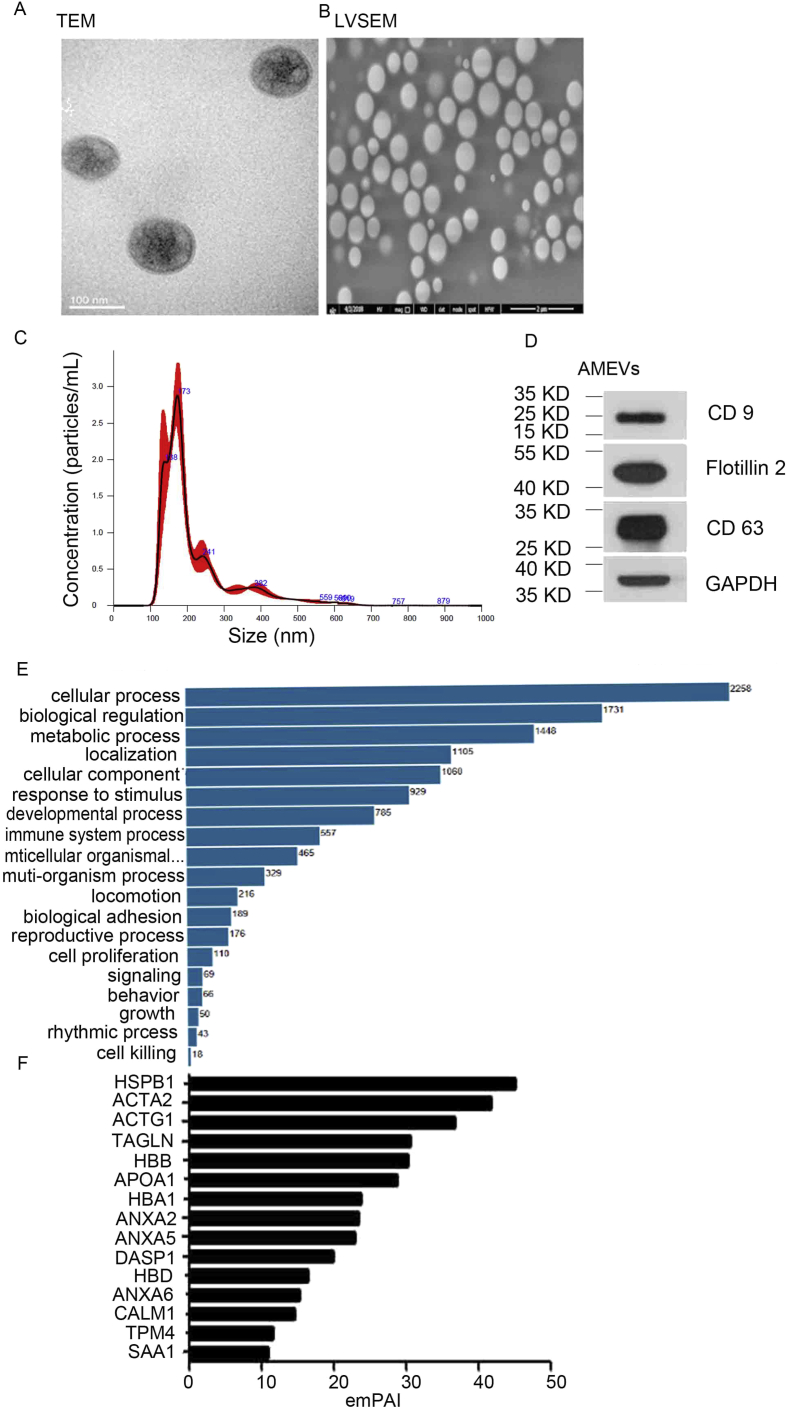
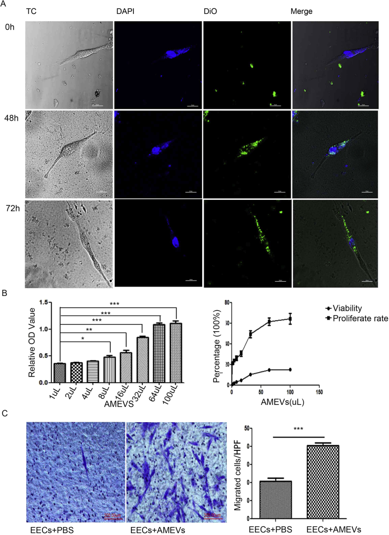
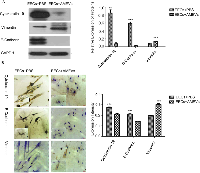
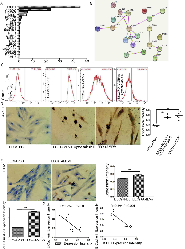
Extracellular vesicles from highly metastatic tumor cells have been shown to mediate epithelial-mesenchymal transition (EMT)-related events in recipient cells. In endometrial epithelial cells, EMT processes are known to be involved in the development of adenomyosis. We aimed to investigate whether adenomyosis-derived extracellular vesicles (AMEVs) are able to induce an EMT process in endometrial epithelial cells. In this study, AMEVs were isolated from patients with adenomyosis and characterized by transmission electron microscopy, Western blot, and nanoparticle tracking. Primary endometrial epithelial cells (EECs) were derived from normal endometrium tissues from patients with leiomyoma and co-cultured with AMEVs . AMEV uptake was examined by fluorescence confocal microscopy. The invasion of EECs was confirmed by Transwell assay. Immunohistochemistry, Western blot, and qRT-PCR were performed on EECs to illustrate the expression levels of cytokeratin 19, E-cadherin, vimentin, and zinc finger E-box-binding homeobox 1 (ZEB1). The results indicated that the cellular fluorescence intensity gradually increased after 48 h of co-culture, but decreased after 72 h. After co-culturing with AMEVs for 72 h, EECs expressed significantly lower levels of cytokeratin 19 and E-cadherin, and significantly higher levels of vimentin and ZEB1. Together these results demonstrated that AMEVs induce an EMT process and enhance the invasion of EECs. These changes may contribute to the pathogenesis and progression of adenomyosis.
来自高转移性肿瘤细胞的细胞外囊泡已被证明可介导受体细胞中与上皮-间质转化(EMT)相关的事件。在子宫内膜上皮细胞中,EMT过程已知参与子宫腺肌病的发展。我们旨在研究子宫腺肌病衍生的细胞外囊泡(AMEV)是否能够诱导子宫内膜上皮细胞发生EMT过程。在本研究中,从子宫腺肌病患者中分离出AMEV,并通过透射电子显微镜、蛋白质免疫印迹和纳米颗粒跟踪进行表征。原代子宫内膜上皮细胞(EEC)来自子宫肌瘤患者的正常子宫内膜组织,并与AMEV共培养。通过荧光共聚焦显微镜检查AMEV的摄取情况。通过Transwell实验确认EEC的侵袭能力。对EEC进行免疫组织化学、蛋白质免疫印迹和qRT-PCR检测,以说明细胞角蛋白-19、E-钙黏蛋白、波形蛋白和锌指E盒结合同源框蛋白1(ZEB1)的表达水平。结果表明,共培养48小时后细胞荧光强度逐渐增加,但72小时后降低。与AMEV共培养72小时后,EEC中细胞角蛋白-19和E-钙黏蛋白的表达水平显著降低,波形蛋白和ZEB1的表达水平显著升高。这些结果共同表明,AMEV可诱导EMT过程并增强EEC的侵袭能力。这些变化可能有助于子宫腺肌病的发病机制和进展。