Westergaard Marie Christine Wulff, Milne Katy, Pedersen Magnus, Hasselager Thomas, Olsen Lars Rønn, Anglesio Michael S, Borch Troels Holz, Kennedy Mia, Briggs Gillian, Ledoux Stacey, Kreuzinger Caroline, Decken Isabel von der, Donia Marco, Castillo-Tong Dan Cacsire, Nelson Brad H, Svane Inge Marie
National Center for Cancer Immune Therapy (CCIT-DK), Department of Oncology, Copenhagen University Hospital, 2730 Herlev, Denmark.
Deeley Research Centre, BC Cancer, Victoria, BC V8R 6V5, Canada.
Cancers (Basel). 2020 Dec 18;12(12):3828. doi: 10.3390/cancers12123828.
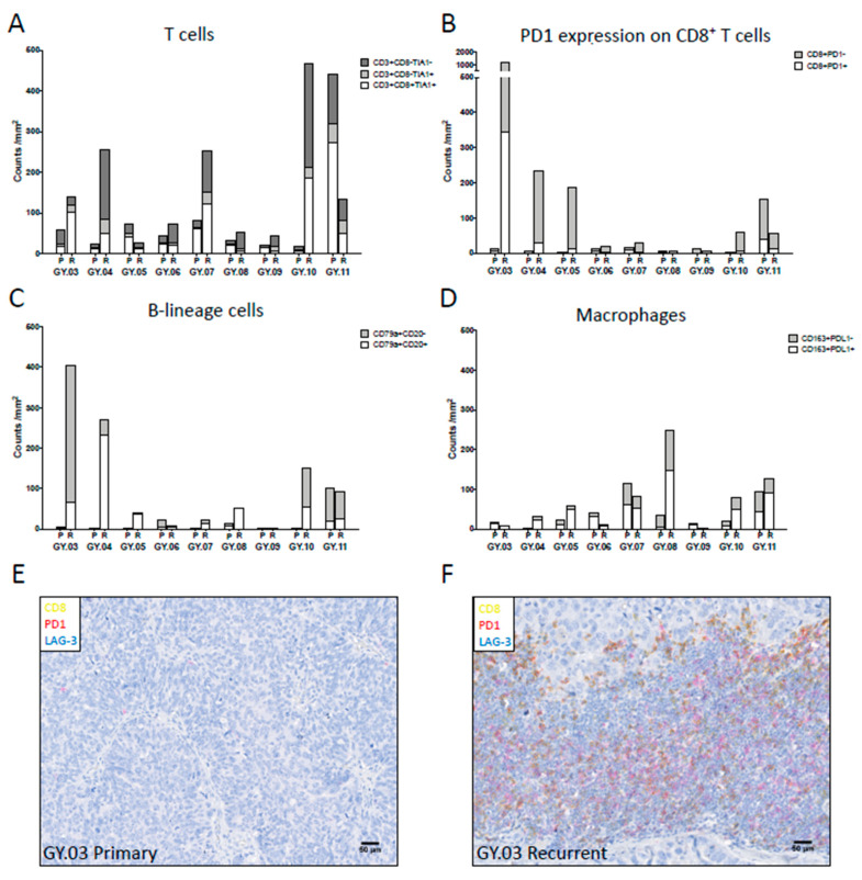
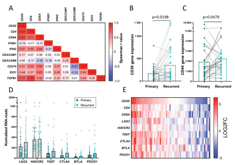
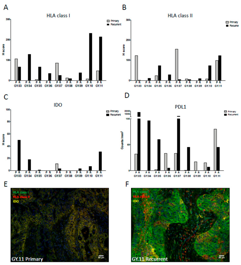
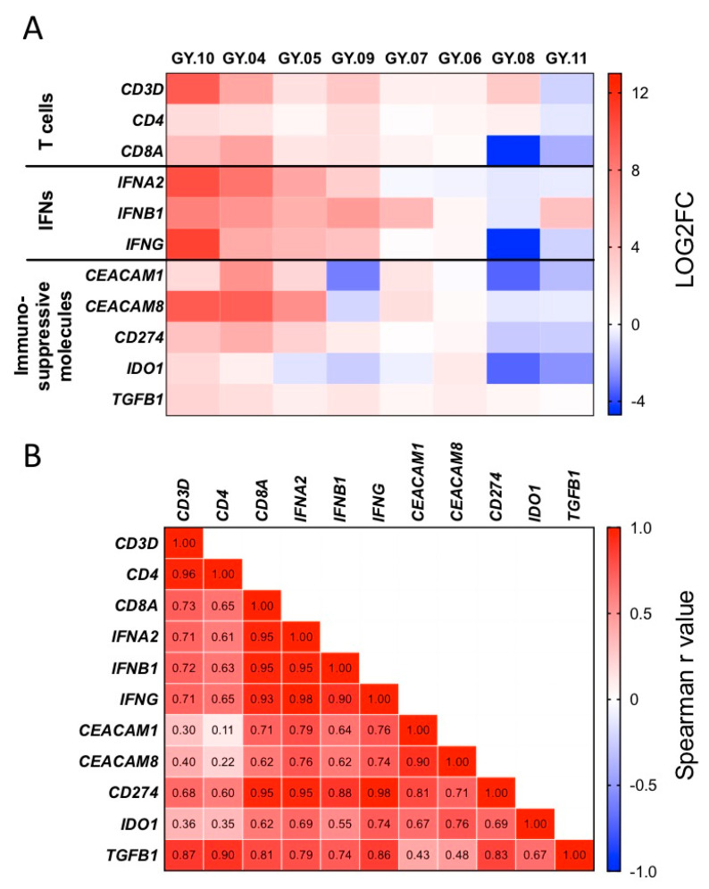
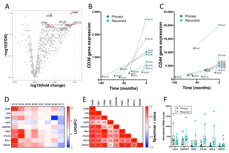
Anti-PD1/PDL1 therapy has proven efficacious against many cancers but only reached modest objective response rates against recurrent ovarian cancer. A deeper understanding of the tumor microenvironment (TME) may reveal other immunosuppressive mechanisms that warrant investigation as immunotherapeutic targets for this challenging disease. Matched primary and recurrent tumors from patients with high-grade serous ovarian carcinoma (HGSC) were analyzed by multicolor immunohistochemistry/immunofluorescence for the presence of T cells, B cells, macrophages, and for the expression of immunosuppressive and HLA molecules. Cancer- and immune-related gene expression was assessed by NanoString analysis. Recurrent tumors showed increased infiltration by immune cells, displayed higher expression of PDL1, IDO, and HLA molecules, and contained more stromal tissue. NanoString analysis demonstrated increased expression of gene signatures related to chemokines and T cell functions in recurrent tumors. The ovarian tumors showed high gene expression of and (TIM3) and enhanced levels of and in recurrent tumors compared to primary tumors. The majority of HGSC developed into a more inflamed phenotype during progression from primary to recurrent disease, including indications of adaptive immune resistance. This suggests that recurrent tumors may be particularly sensitive to inhibition of adaptive immune resistance mechanisms.
抗PD1/PDL1疗法已被证明对多种癌症有效,但对复发性卵巢癌仅达到适度的客观缓解率。对肿瘤微环境(TME)的更深入了解可能会揭示其他免疫抑制机制,这些机制值得作为这种具有挑战性疾病的免疫治疗靶点进行研究。通过多色免疫组织化学/免疫荧光分析高级别浆液性卵巢癌(HGSC)患者匹配的原发性和复发性肿瘤中T细胞、B细胞、巨噬细胞的存在情况以及免疫抑制分子和HLA分子的表达。通过NanoString分析评估癌症和免疫相关基因的表达。复发性肿瘤显示免疫细胞浸润增加,PDL1、IDO和HLA分子表达更高,且含有更多的基质组织。NanoString分析表明,复发性肿瘤中与趋化因子和T细胞功能相关的基因特征表达增加。与原发性肿瘤相比,卵巢肿瘤显示 和T细胞免疫球蛋白黏蛋白3(TIM3)的基因表达较高,且复发性肿瘤中 和 的水平升高。在从原发性疾病进展到复发性疾病的过程中,大多数HGSC发展为更具炎症性的表型,包括适应性免疫抵抗的迹象。这表明复发性肿瘤可能对适应性免疫抵抗机制的抑制特别敏感。