Suppr超能文献

也门人群样本中牙髓石患病率的影像学评估

Radiographic Assessment of the Prevalence of Pulp Stones in a Yemeni Population Sample.

作者信息

Kalaji Mohamed Nader, Habib Adnan Asaad, Alwessabi Mohamed

机构信息

Department of Endodontics, School of Dentistry, University of Oslo, Oslo, Norway.

Department of Restorative Dental Sciences, AL-Farabi Colleges of Dentistry and Nursing, Saudi Arabia.

出版信息

Eur Endod J. 2017 Oct 10;2(1):1-6. doi: 10.14744/eej.2017.17024. eCollection 2017.

Abstract

OBJECTIVE

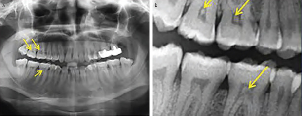
To determine the prevalence and distribution of pulp stones in the posterior teeth of a sample of adult Yemeni dental patients using digital panoramic radiographs.

METHODS

In total, 913 panoramic radiographs from patients attending the hospital dental clinics of at University of Sciences and Technology, Sana'a, Yemen, from January 2013 to December 2014 were examined. The occurrence of pulp stones in the posterior teeth of adult subjects was recorded. Associations between pulp stones and gender, age, arch, side and tooth type were studied.

RESULTS

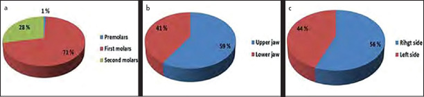
The overall prevalence of pulp stones was 18.6% for individuals (170 out of 913 subjects) and 3.99% for examined teeth (351 out of 8802 teeth). The pulp stone occurrence was significantly higher in the maxilla than in the mandible for each tooth type and location (P<0.001). Pulp stones occurred more often on the right side (P<0.001). First molars represented 71% of the affected teeth with the maxillary right first molar showing the highest occurrence. Fifty-six percent of the affected subjects had pulp stones in more than one tooth. No significant difference in the occurrence of pulp stones was detected between genders or among age groups (P>0.05).

CONCLUSION

The prevalence of pulp stones is different among populations. Pulp stones were found in approximately one-fifth of subjects in the Yemeni population, where up to 90% of the population have a Qat-chewing habit. This habit usually causes mechanical and chemical irritation and results in pulp calcification.

摘要

目的

使用数字化全景X线片确定也门成年牙科患者样本后牙中髓石的患病率及分布情况。

方法

对2013年1月至2014年12月期间也门萨那科学与技术大学医院牙科诊所患者的913张全景X线片进行检查。记录成年受试者后牙中髓石的发生情况。研究髓石与性别、年龄、牙弓、侧别和牙型之间的关联。

结果

个体髓石总体患病率为18.6%(913名受试者中有170人),检查牙齿的患病率为3.99%(8802颗牙齿中有351颗)。每种牙型和位置的髓石发生率在上颌明显高于下颌(P<0.001)。髓石在右侧更常见(P<0.001)。第一磨牙占患牙的71%,其中上颌右侧第一磨牙发生率最高。56%的患牙受试者有多颗牙齿出现髓石。性别和年龄组之间髓石发生率无显著差异(P>0.05)。

结论

不同人群中髓石的患病率不同。在也门人群中,约五分之一的受试者发现有髓石,该国高达90%的人口有咀嚼巧茶的习惯。这种习惯通常会引起机械和化学刺激,导致牙髓钙化。

https://cdn.ncbi.nlm.nih.gov/pmc/blobs/c9a5/7757961/4ae9f3316df0/EEJ-2-25-g001.jpg

相似文献

1
Radiographic Assessment of the Prevalence of Pulp Stones in a Yemeni Population Sample.
Eur Endod J. 2017 Oct 10;2(1):1-6. doi: 10.14744/eej.2017.17024. eCollection 2017.
2
Detecting calcified pulp stones in patients with periodontal diseases using digital panoramic and periapical radiographies.
J Dent Sci. 2022 Apr;17(2):965-972. doi: 10.1016/j.jds.2021.12.010. Epub 2022 Jan 11.
4
Dental pulp stone formation during orthodontic treatment: A retrospective clinical follow-up study.
Niger J Clin Pract. 2017 Jan;20(1):37-42. doi: 10.4103/1119-3077.164357.
7
A radiographic assessment of the prevalence of pulp stones in Australians.
Aust Dent J. 2002 Mar;47(1):36-40. doi: 10.1111/j.1834-7819.2002.tb00301.x.
9
The prevalence of pulp stones in a Turkish population. A radiographic survey.
Med Oral Patol Oral Cir Bucal. 2012 Mar 1;17(2):e212-7. doi: 10.4317/medoral.17400.
10
Radiographic assessment of the prevalence of pulp stones in Malaysians.
J Endod. 2015 Mar;41(3):333-7. doi: 10.1016/j.joen.2014.10.015. Epub 2014 Dec 2.

引用本文的文献

5
Early diagnosis of aortic calcification through dental X-ray examination for dental pulp stones.
Sci Rep. 2023 Oct 30;13(1):18576. doi: 10.1038/s41598-023-45902-w.
6
The prevalence of pulp stones in a Turkish paediatric cohort from the Isparta district: an 8-year retrospective radiographic study.
Eur Arch Paediatr Dent. 2023 Dec;24(6):729-736. doi: 10.1007/s40368-023-00836-9. Epub 2023 Sep 5.
7
Detecting calcified pulp stones in patients with periodontal diseases using digital panoramic and periapical radiographies.
J Dent Sci. 2022 Apr;17(2):965-972. doi: 10.1016/j.jds.2021.12.010. Epub 2022 Jan 11.
8
Incidence and distribution of pulp stones among Southern Saudi Arabian sub-population.
SAGE Open Med. 2021 Dec 23;9:20503121211062796. doi: 10.1177/20503121211062796. eCollection 2021.

本文引用的文献

1
Assessing pulp stones by cone-beam computed tomography.
Clin Oral Investig. 2017 Sep;21(7):2327-2333. doi: 10.1007/s00784-016-2027-5. Epub 2016 Dec 9.
2
Radiographic assessment of the prevalence of pulp stones in Malaysians.
J Endod. 2015 Mar;41(3):333-7. doi: 10.1016/j.joen.2014.10.015. Epub 2014 Dec 2.
4
Can dental pulp calcification predict the risk of ischemic cardiovascular disease?
J Dent (Tehran). 2013 Sep;10(5):456-60. Epub 2013 Sep 30.
6
Assessment of the prevalence of pulp stones in a sample of Turkish Central Anatolian population.
ScientificWorldJournal. 2012;2012:804278. doi: 10.1100/2012/804278. Epub 2012 May 3.
7
The prevalence of pulp stones in a Turkish population. A radiographic survey.
Med Oral Patol Oral Cir Bucal. 2012 Mar 1;17(2):e212-7. doi: 10.4317/medoral.17400.
9
Association between dental pulp stones and calcifying nanoparticles.
Int J Nanomedicine. 2011 Jan 7;6:109-18. doi: 10.2147/IJN.S13267.
10
A radiographic correlation between systemic disorders and pulp stones.
Indian J Dent Res. 2010 Jul-Sep;21(3):369-73. doi: 10.4103/0970-9290.70806.

文献AI研究员

20分钟写一篇综述,助力文献阅读效率提升50倍。

立即体验

用中文搜PubMed

大模型驱动的PubMed中文搜索引擎

马上搜索

文档翻译

学术文献翻译模型,支持多种主流文档格式。

立即体验