Department of Psychiatry, School of Clinical Medicine, University of Cambridge, Level E4 Cambridge Biomedical Campus, Cambridge, CB2 0SP, UK.
Centre for Dementia Prevention, University of Edinburgh Centre for Clinical Brain Sciences, Edinburgh, UK.
J Neurol. 2021 May;268(5):1962-1971. doi: 10.1007/s00415-020-10383-8. Epub 2021 Jan 9.
Structural brain changes associated with Alzheimer's disease (AD) can occur decades before the onset of symptoms. The Cardiovascular Risk Factors, Aging, and Dementia (CAIDE) score has been suggested to be associated with accelerated brain atrophy in middle-aged subjects but the regional specificity of atrophic areas remains to be elucidated.
3T T1-weighted magnetic resonance imaging scans of 160 cognitively healthy middle-aged participants (mean age = 52) in the PREVENT-Dementia cohort, from baseline and from follow-up after 2 years, were examined. Images were preprocessed using Computational Anatomy Toolbox 12. Voxel-based morphometry was performed in FSL 6.0.1 to identify areas of grey matter (GM) volume differences both cross-sectionally and longitudinally between subjects with high and low baseline CAIDE score (CAIDE score was dichotomized at cohort-median). A GM percentage of change map was created for each subject for evaluation of atrophy over 2 years. Analyses were adjusted for age, gender, education and total intracranial volume.
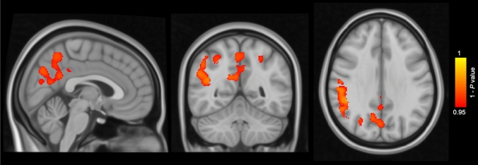
Compared to subjects with CAIDE score ≤ 6 (low risk), subjects with CAIDE score > 6 (high risk) showed lower GM volume in the temporal, occipital, and fusiform cortex and lingual gyrus at baseline, and greater percentage of GM loss over 2 years in the supramarginal gyrus, angular gyrus, precuneus, lateral occipital cortex, superior parietal lobule and cingulate gyrus (corrected P < 0.05).
This study demonstrated accelerated GM atrophy concentrated in several AD signature cortical regions in healthy middle-aged subjects with high CAIDE scores.
与阿尔茨海默病(AD)相关的结构性脑变化可在症状出现前数十年发生。心血管危险因素、衰老和痴呆(CAIDE)评分被认为与中年受试者的脑萎缩加速有关,但萎缩区域的局部特异性仍有待阐明。
在 PREVENT-Dementia 队列中,对 160 名认知健康的中年参与者(平均年龄=52 岁)的 3T T1 加权磁共振成像扫描进行了检查,这些参与者在基线和 2 年后的随访中接受了检查。使用 Computational Anatomy Toolbox 12 对图像进行预处理。在 FSL 6.0.1 中进行基于体素的形态测量,以识别高基线 CAIDE 评分(CAIDE 评分在队列中位数处分为二分类)和低基线 CAIDE 评分受试者之间的灰质(GM)体积差异。为每个受试者创建 GM 百分比变化图,以评估 2 年内的萎缩情况。分析调整了年龄、性别、教育和总颅内体积。
与 CAIDE 评分≤6(低风险)的受试者相比,CAIDE 评分>6(高风险)的受试者在基线时颞叶、枕叶和梭状回以及舌回的 GM 体积较低,2 年内 GM 损失的百分比更高在缘上回、角回、楔前叶、外侧枕叶、顶下小叶和扣带回(校正 P<0.05)。
本研究表明,在 CAIDE 评分较高的健康中年受试者中,AD 特征性皮质区域集中存在加速 GM 萎缩。