Division of ENT Diseases, Department of Clinical Sciences, Intervention and Technology, Karolinska Institutet, Stockholm, Sweden.
Department of Otorhinolaryngology, Karolinska University Hospital, Stockholm, Sweden.
Cancer Sci. 2021 Mar;112(3):1048-1059. doi: 10.1111/cas.14816. Epub 2021 Feb 15.
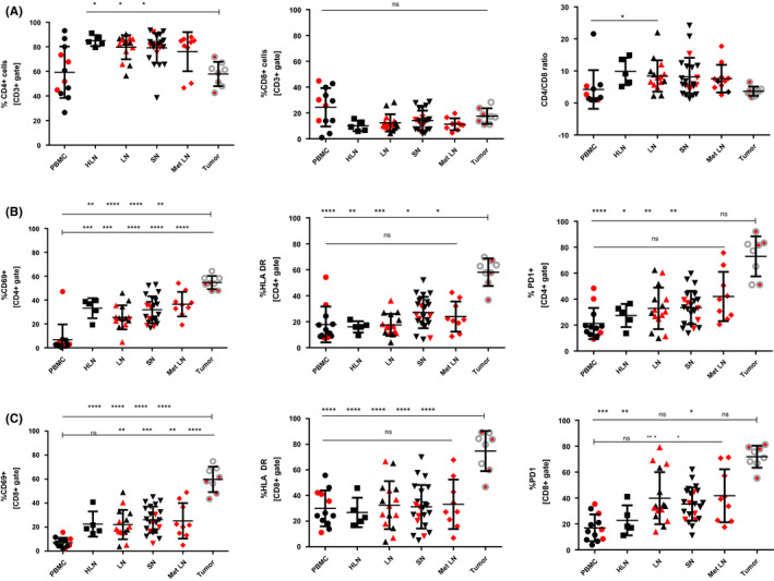
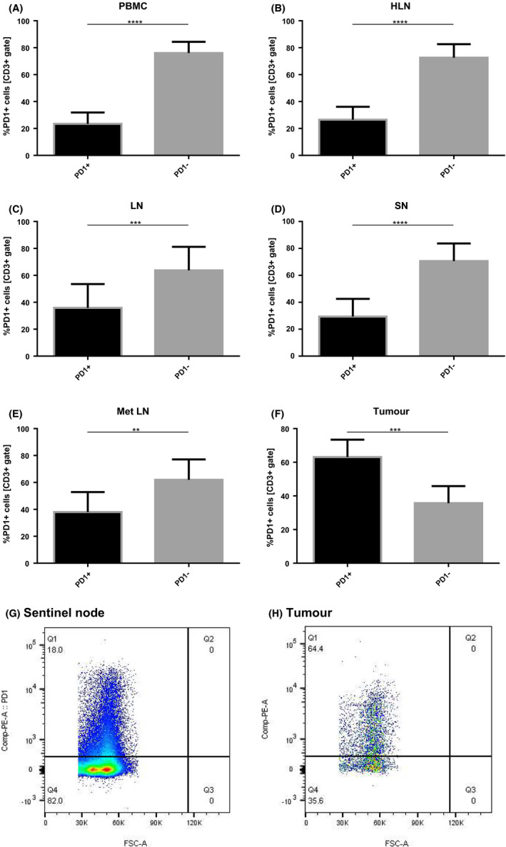
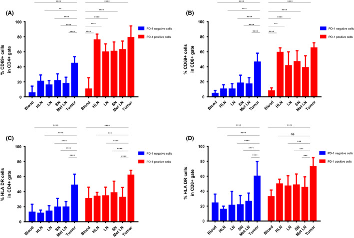
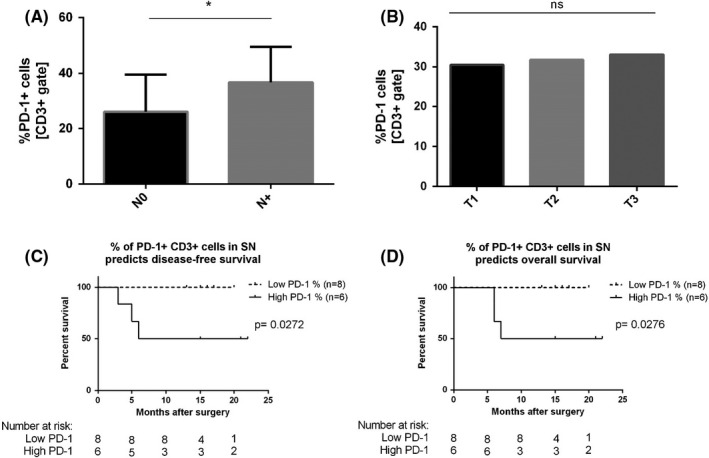
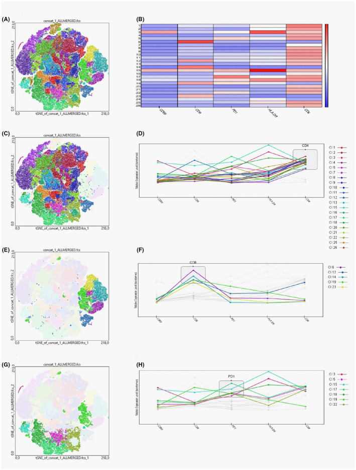
Anticancer immunotherapies have revolutionized cancer management, yet the effect of systemic anti-programmed cell death protein 1 (PD-1) treatment is predominantly studied in tumor-infiltrating lymphocytes (TILs). Its impact on PD-1 expressing cells in tumor-draining lymph nodes (TDLNs) is not well understood and yet to be explored. Thus, further research aiming for better understanding of the PD-1 pathway not only in tumor tissue but also in TDLNs is warranted. In this study, we investigated the expression of PD-1, CD69, and HLA-DR on CD4 and CD8 T cells by flow cytometry analysis of peripheral blood mononuclear cells (PBMCs), TDLNs, and tumor samples from patients with oral squamous cell carcinoma (OSCC). Our data showed that both helper and cytotoxic T lymphocytes in OSCC tissue were highly activated and expressed high level of PD-1 (over 70% positivity). Lymphocytes in TDLNs and peripheral blood expressed significantly lower levels of PD-1 and other activation markers compared to TILs. Moreover, we demonstrated that a significant fraction of PD-1 negative TILs expressed high levels of human leukocyte antigen - DR isotype and CD69. In contrast, PD-1 negative cells in TDLNs and PBMCs scarcely expressed the aforementioned activation markers. Furthermore, we proved that patients with a high percentage of CD3 PD-1 cells in tumor-draining lymph nodes had significantly lower disease-free and overall survival rates (log-rank test P = .0272 and P = .0276, respectively). Taken together, we proved that flow cytometry of lymph nodes in OSCC is feasible and may be used to investigate whether PD-1 levels in TDLNs correspond with survival and potentially with response to anti-PD-1 therapy. Such knowledge may ultimately help guide anti-PD-1 treatment.
癌症免疫疗法已经彻底改变了癌症的治疗方式,但系统抗程序性细胞死亡蛋白 1(PD-1)治疗的效果主要在肿瘤浸润淋巴细胞(TIL)中进行研究。其对肿瘤引流淋巴结(TDLNs)中 PD-1 表达细胞的影响尚未得到很好的理解,也有待探索。因此,为了更好地理解 PD-1 通路,不仅在肿瘤组织中,而且在 TDLNs 中进行进一步的研究是有必要的。在这项研究中,我们通过流式细胞术分析外周血单核细胞(PBMCs)、TDLNs 和口腔鳞状细胞癌(OSCC)患者的肿瘤样本,研究了 PD-1、CD69 和 HLA-DR 在 CD4 和 CD8 T 细胞上的表达。我们的数据表明,OSCC 组织中的辅助性和细胞毒性 T 淋巴细胞均高度激活,并表达高水平的 PD-1(超过 70%的阳性率)。TDLNs 和外周血中的淋巴细胞表达的 PD-1 和其他激活标志物明显低于 TILs。此外,我们证明了相当一部分 PD-1 阴性 TILs 表达高水平的人类白细胞抗原-DR 同工型和 CD69。相比之下,TDLNs 和 PBMCs 中 PD-1 阴性细胞很少表达上述激活标志物。此外,我们证明了肿瘤引流淋巴结中 CD3 PD-1 细胞百分比高的患者无病生存率和总生存率明显降低(对数秩检验 P =.0272 和 P =.0276)。综上所述,我们证明了 OSCC 淋巴结的流式细胞术是可行的,并且可以用于研究 TDLNs 中的 PD-1 水平是否与生存相关,并且可能与抗 PD-1 治疗的反应相关。这些知识最终可能有助于指导抗 PD-1 治疗。