Department of Orthopaedics and Trauma Surgery, Universitätsklinikum Bonn, Venusberg-Campus 1, 53127, Bonn, Germany.
U.O.C. 1° Clinica Ortopedica, ASST Centro Specialistico Ortopedico Traumatologico Gaetano Pini-CTO, Piazza Cardinal Ferrari 1, Milan, 20122, Italy.
Arch Orthop Trauma Surg. 2022 May;142(5):813-821. doi: 10.1007/s00402-021-03753-y. Epub 2021 Jan 23.
Preventing nerve injury is critical in elbow surgery. Distal extension of medial approaches, required for coronoid fracture fixation and graft-replacement, may endanger the median nerve. This study aims to describe an easily identifiable and reproducible anatomical landmark to localize the median nerve distal to the joint line and to delineate how its relative position changes with elbow flexion and forearm rotation.
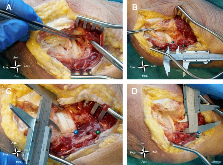
The median nerve and the ulnar insertion of the brachialis muscle were identified in eleven fresh-frozen cadaveric specimens after dissection over an extended medial approach. The elbow was brought first in full extension and then in 90° flexion, and the shortest distance between the two structures was measured while rotating the forearm in full pronation, neutral position and full supination.
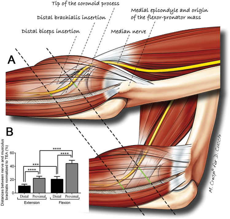
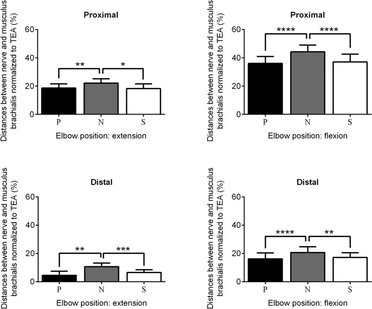
The distance between the median nerve and the brachialis insertion was highest with the elbow flexed and the forearm in neutral position. All distances measured in flexion were larger than those in extension, and all distances measured from the most proximal point of the brachialis insertion were larger than those from the most distal point. Distances in pronation and in supination were smaller than to those in neutral forearm position.
The ulnar insertion of the brachialis is a reliable landmark to localize and protect the median nerve at the level of the coronoid base. Elbow flexion and neutral forearm position increase significantly the safety margins between the two structures; this information suggests some modifications to the previously described medial elbow approaches.
Basic Science Study.
预防神经损伤在肘部手术中至关重要。为了固定冠状突骨折和进行移植物置换,内侧入路需要向远侧延伸,这可能会危及正中神经。本研究旨在描述一个易于识别和可重复的解剖学标志,以确定位于关节线以下的正中神经位置,并阐明其相对位置随肘弯曲和前臂旋转而发生的变化。
在通过扩展的内侧入路进行解剖后,在十一个新鲜冷冻的尸体标本中识别正中神经和肱二头肌的尺骨插入部。首先将肘部置于完全伸展状态,然后置于 90°弯曲状态,同时在前臂完全旋前、中立位和完全旋后时测量这两个结构之间的最短距离。
当肘部弯曲且前臂处于中立位时,正中神经和肱二头肌插入部之间的距离最高。所有在弯曲时测量的距离均大于伸展时的距离,并且所有从肱二头肌插入部最近端测量的距离均大于从最远端测量的距离。在旋前和旋后时的距离小于中立位时的距离。
肱二头肌的尺骨插入部是在冠状突基部定位和保护正中神经的可靠标志。肘弯曲和中立位前臂显著增加了这两个结构之间的安全距离;这些信息提示对先前描述的内侧肘部入路进行一些修改。
基础科学研究。